The patient has acute pain in the ankle region. The ankle often swells up quickly and develops bruises. The patient can no longer walk without pain.
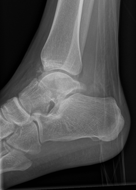
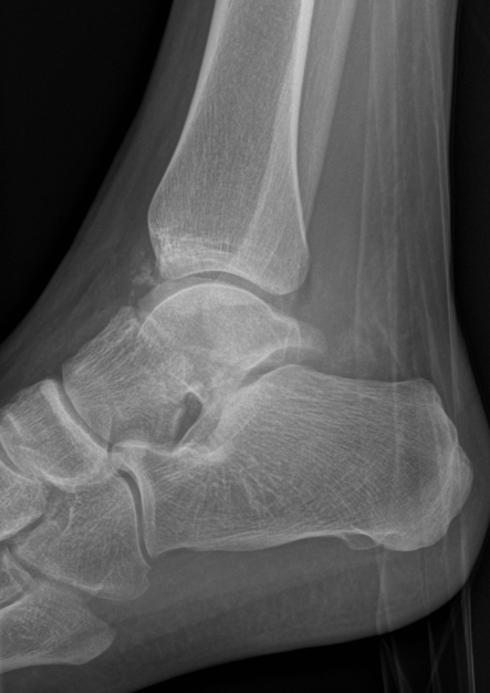
Diagnosing an ankle (talus) fracture
Due to the anatomical proximity of nerves and blood vessels, it is vital that the motor/sensory function and blood supply to the patient’s foot are examined as soon as possible. The method of choice for diagnosing the problem consists of x-rays in 2 planes. A CT scan is also indicated in many cases. This can show the exact position of the bone fragments and is helpful when planning the operation. An MRI scan may be necessary in rare cases (most often to ascertain damage to the ankle caused by poor blood circulation, a condition known as osteochondritis dissecans).
Treating a broken ankle (talus fracture)
Fractures that are not displaced and have no widely dispersed fragments can be treated conservatively. The patient’s lower leg is first immobilised in a splint. The ankle must be elevated and cooled until the swelling in the injured area subsides. The splint is then exchanged for a circular plaster cast. After another 4 weeks, this cast can be removed and a walking cast applied. The leg will not be able to bear the patient’s full weight until 8 weeks have passed. However, this only applies to Hawkins type I injuries.
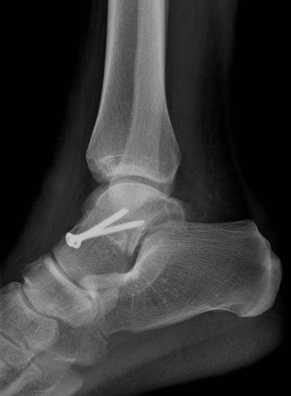
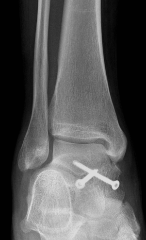
Fractures from type II upwards and breaks with dispersed fragments must be treated surgically. The standard surgical procedure involves the insertion of small screws. It may be possible to realign the ankle or fragments without cutting through the skin. If not, an incision must be made to realign the ankle and insert implants (screws) to hold the broken bones in place.
Talus fracture Hawkins type I-II
Anatomical repositioning and 3.5 mm lag screw osteosynthesis
Post-surgical treatment of ankle (talus) fractures
After the surgery, the patient will have to wear a plaster splint until the swelling goes down and the wound heals. No weight should be put on the operated limb for at least 6 weeks (up to 12 weeks depending on how well the ankle heals). The patient will therefore have to use crutches to move about. Physiotherapy incorporating exercises to improve mobility in the upper and lower ankle joint are advisable during this period. If the talus has lost all contact with the upper and lower ankle joint (total dislocation), it must be reset without delay in order to minimise the risk of the bone tissue dying due to insufficient blood supply (avascular necrosis). This is accomplished by applying traction with the knee bent. Here, too, early passive mobilisation following the bone resetting is decisive.
Possible complications / prognosis (broken ankle)
The prognosis is largely determined by the necrosis rate, i.e. the percentage of bone cells that die as the result of impaired blood circulation. For Hawkins type I fractures, the rate is 5 to 10%, for Hawkins type II 40% to 50%, and for Hawkins types III and IV up to 100%. Not all necrosis of the talus leads to arthrosis (wear and tear) that requires treatment. The incidence of arthrosis for each Hawkins type is 20%, 50% and 75% respectively. For this reason, surgery must be performed without delay so that the talus can be restored and realigned with the other bones in the ankle joint. Fusion surgery is often required if arthrosis develops later on.
For patients
As a patient, you cannot register for a consultation directly. Please ask your family doctor, your specialist to refer you. Please use our contact form if you have any questions.