Treatment of pelvic injuries / pelvic ring fractures

Fractures of the pelvic ring account for approx. 4 percent of all fractures. This injury is found in 25 percent of patients who suffer serious accidents (high-energy impact injuries: road accidents, falls from great heights).

Bleeding occurring in the vicinity of unstable pelvic ring fractures may be difficult to control. It can be so profuse that the patient’s life is in grave danger. In these cases, the pelvis must be stabilised immediately. The urinary tract is also at risk due to its proximity to the site of the injury. Older patients frequently suffer so-called “stable fractures” (more than 50% of pelvic fractures) as a result of a fall (low-energy trauma).

Links: 3D-Darstellung einer Computertomographie nach kombiniertem linksseitigem Becken- und Hüftpfannenbrauch. Rechts:Postoperatives Röntgen nach Fixation mit Platten und Schrauben.

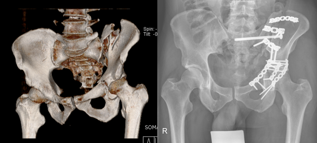
Left: 3D computer tomography following combined fracture of the pelvis and acetabulum. Right: Postoperative x-ray following fixation with plates and screws.

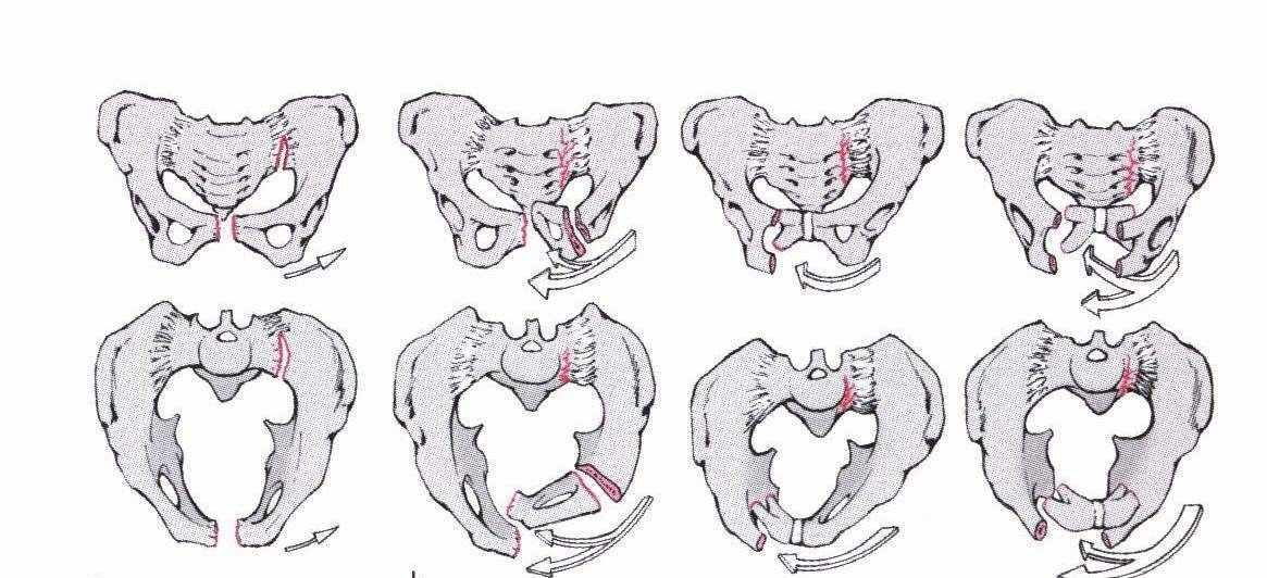
The forces involved are:

  • Lateral compression
  • Anterior-posterior compression
  • Vertical shear (along the spinal axis)
  • Combined injuries
Einwirkende Kräfte und resultierende Brüche des Beckens

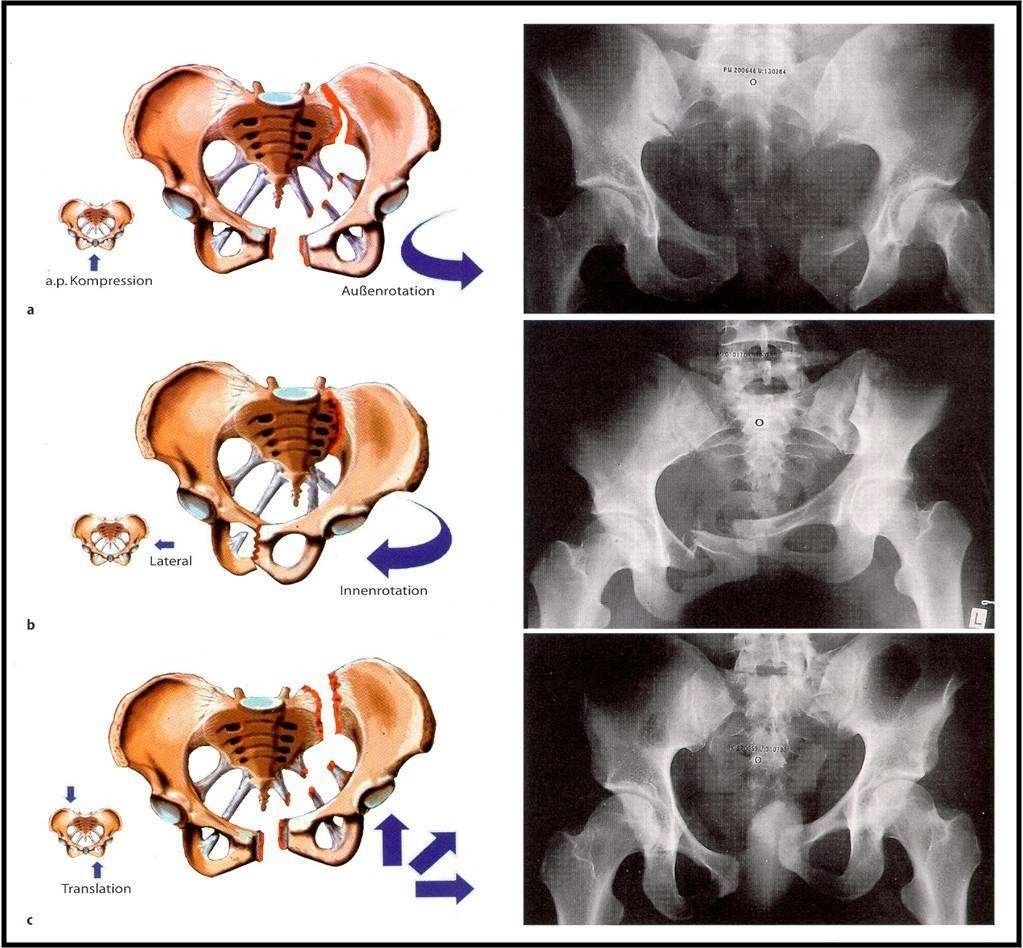
Forces acting on the pelvis and resulting fractures (Orthopädie und Unfallchirurgie up2date [Orthopaedics and Trauma Surgery up2date], Thieme)

 Grundsätzlich muss man zwischen stabilen und instabilen Beckenbrüchen unterscheiden. Wichtig: Das Becken ist dann stabil, wenn die wesentlichen Strukturen des hinteren Beckenrings unerletzt sind. Das Becken ist dann instabil, wenn knöcherne oder Bandstrukturen im Bereich des vorderen und hinteren Becknringes verletzt sind. Es gibt verschiedene Klassifikationssysteme, die aber letztendlich alle zwischen stabil, rotatorisch instabil und rotatorisch/vertikal instabil unterscheiden. Bei den stabilen Verletzungen handelt sich um Brüche oder Abrisse an den Darmbeinschauflen, dem Sitzbein, Schambein oder dem Steissbein unterhalb der Gelenkverbindungen, die die Stabilität des Beckenringhs nicht gefährden. Diese Verletzungen können häufig ohne eine Operation behandelt werden. Hier stehen die Schmerzbekämfungen und die Mobilisation des Patienten im Vordergrund. BildBeckenringRotationsinstabilitaet.jpgAbbildung: Stabile Beckenbrüche (Ficklscherer, Orthopädie und Traumatologie, Urban & Fischer) Rotationsinstabilität besteht beispielsweise bei Sprengungen der vorderen Beckenanteile oder aber auch Verschiebungen der hinteren Strukturen. Hier ist aber die vertikale Stabilität erhalten. Bei aussenrotierter Beckenhälfte spricht man von einer Open-book-Verletzung (siehe Abbildung 5). Diese Brüche werden je nach Verletzungsausmass konservativ oder operativ behandelt. BeckenringfrakturenAbb4.jpg Abbildung: Rotatorisch instabile Brüche (Ficklscherer. Orthopädie und Traumatologie, Urban & Fischer)​ Beckenfringfrakturen_Abb5Open_book.jpgAbbildung: Open-book-Verletzung Die Instabilsten Verletzungen stellen die Kombinationsverletzungen dar. Häufig sind sie mit anderen Verletzungen des Skelettsystems und anderer Körperregionen kombiniert. Hierbei sind gleichzeitig Strukturen des vorderen und des hinteren Beckenrings beteiligt. Die rotatorisch und vertikal instabile Brüche stellen eine absolute Operationsindikation dar. Je nach Zustand des Verletzten wird vor der definitiven Versorgung (häufig Platten oder Schrauben) eine notfallmässige externe Stabilisierung mittels Fixateur externe oder interne vorgenommen. Bild Becken 3.pngAbbildung: Links: Computertomographie nach Ausbruchverletzung des ersten Kreuzbeinwirbels. Rechts: Postoperatives Röntgen nach minimal-invasiver Stabilisation mittels langer Schrauben über kleine Hautschnitte.

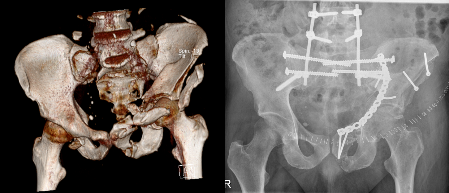
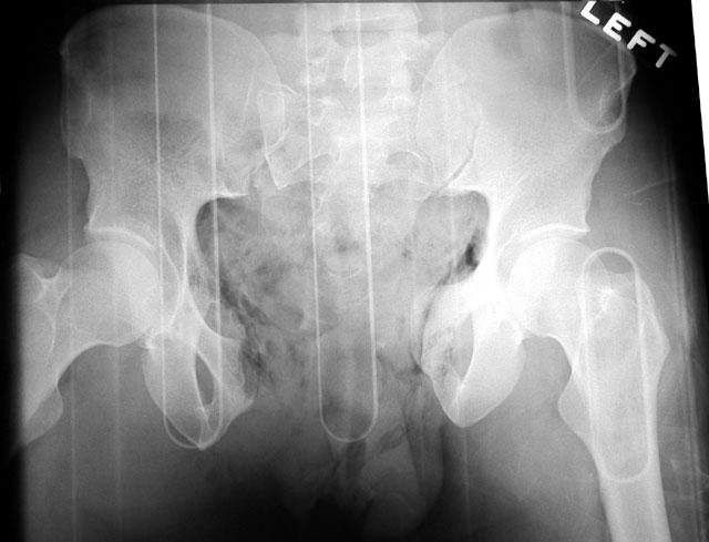
Left: 3D computed tomography imaging following an extremely severe bilateral combined fracture of the pelvis and acetabulum with separation of the pelvis and lumbar spine. Right: Postoperative x-ray following realignment of hips and pelvic ring and reconnection with lumbar spine using plates, screws and rods.

A distinction must always be made between stable and unstable pelvic fractures. Important:

  • The pelvis is stable when the major structures in the posterior pelvic ring are uninjured.
  • The pelvis is unstable when bony or ligamentous structures in the anterior and posterior pelvic ring have been injured.

There are various classification systems; ultimately, however, they all distinguish between stable, rotationally unstable and rotationally/vertically unstable.

Stable injuries include fractures or avulsion of the iliac wings, ischium, pubis or coccyx below the articulated joints which do not affect the stability of the pelvic ring. These injuries can often be treated without surgery. The priorities in such cases are pain management and patient mobilisation.

Illustration Stabile Beckenbrüche

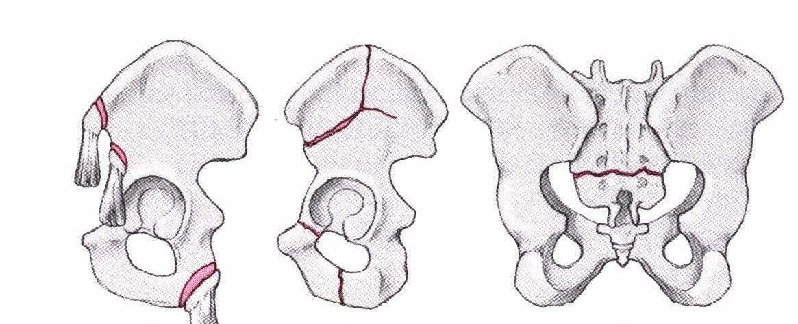
Stable pelvic fractures (Ficklscherer, Orthopädie und Traumatologie [Orthopaedics and Traumatology], Urban & Fischer)

Rotational instability exists for example when the anterior parts of the pelvis have been dislocated or the posterior structures displaced. However, the vertical stability of the pelvis is not affected. External rotation of one side of the pelvis is referred to as an open book injury (cf. fig. 5). These fractures are treated conservatively or surgically depending on the extent of the injury.

Illustration Rotatorisch instabile Brüche

Rotationally unstable fractures (Ficklscherer, Orthopädie und Traumatologie, Urban & Fischer)​

Röntgenbild open book-Verletzung

Combined injuries are the most unstable of all. They are frequently combined with additional injuries to the skeletal system and other body parts. The anterior and posterior structures of the pelvic ring are both affected. Surgery is essential in the case of rotationally and vertically unstable fractures. Depending on the injured patient’s condition, emergency external stabilisation is performed using an external or internal fixator before the definitive treatment is administered (commonly plates or screws).

Links: Computertomographie nach Ausbruchverletzung des ersten Kreuzbeinwirbels. Rechts: Postoperatives Röntgen nach minimal-invasiver Stabilisation mittels langer Schrauben über kleine Hautschnitte.

Open book injury

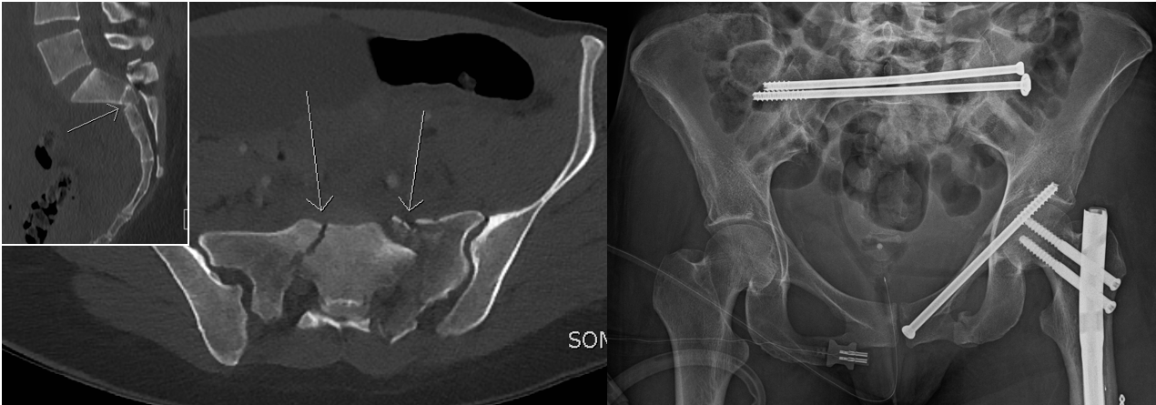
Left: Computed tomography following avulsion injury of the first sacral vertebrum. Right: Postoperative x-ray following minimally invasive stabilisation using long screws inserted through small incisions in the skin.

For patients

As a patient you cannot register for a consultation directly. Please ask your family doctor or your specialist to refer you.

contact form

For assigners

University Hospital Zurich
Department of Traumatology
Rämistrasse 100
8091 Zurich

Tel. +41 44 255 27 55
assign online

Responsible Department