The regional distribution of the tracer in the myocardium makes it possible to visualize ischemia or scarring of the left ventricular myocardium. In addition, the extent and exact localization of myocardial ischemia allow an optimal treatment strategy to be defined for each patient. The PET examination offers a higher resolution with lower radiation exposure than SPECT.
Cardiac MRI also makes it possible to visualize myocardial perfusion, but a contrast agent containing gadolinium is used instead of the radioactive tracer. Finally, stress echocardiography can also detect circulatory disorders indirectly through wall motion disturbances that occur under stress.
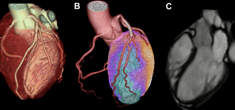
Complementary to SPECT and PET, computed tomography (CT) coronary angiography does not provide any information on myocardial blood flow, but does show wall changes or stenoses of the coronary arteries. The spatial resolution of the coronary CT has been continuously improved (currently 0.23 mm) and the radiation exposure reduced (currently 0.6 mSv). The strength of cardiac CT lies in its negative predictive value.
SPECT
- Advantages: Diagnostic and prognostic accuracy. Long experience. Short scanning time.
- Disadvantages: Radiation exposure (3-9 mSv)
PET
- Advantages: Very high diagnostic accuracy. Ischemia and microcirculatory disorder (NH3) and vitality assessment (FDG).
- Disadvantages: Radiation exposure (2-4 mSv)
Since the end of 2014, the latest technological innovation has been available, namely an integrated PET/MR, which comprises both a PET with state-of-the-art detector technology and a 3T MRI device. In addition to its use in neurology and oncology, combined PET/MR hybrid diagnostics is also being introduced. As with PET/CT hybrid diagnostics, PET/MR allows the diagnostic significance of ischemia clarification, vitality diagnostics, cardiomyopathies and sarcoidosis to be further increased.