What is a mammogram?
A mammogram is an X-ray examination of the breast that allows us to detect benign and malignant changes. These include, for example, fibroadenomas, lipomas, mastopathy, microcalcifications—which may indicate precancerous breast lesions—or breast cancer (mammary carcinoma) itself. Cysts, which are also common in the breasts of many women, are, however, more easily detected during a breast cancer ultrasound (sonography).
In conventional mammography, the breasts are firmly compressed one at a time between two Plexiglas plates. Then, medical radiology technicians (MTRA) take images of each breast from two different angles. This results in a total of four black-and-white X-ray images—two of each breast. We then interpret these findings and classify them as normal or suspicious.
Mammography Without Compression: Breast CT
The USZ is the first institute in the world to have a spiral computed tomography scanner for the female breast. This device performs mammograms without the compression that was previously required. This means the patient no longer needs to be afraid of the examination, which has sometimes been painful in the past.
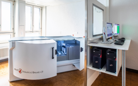
We often combine mammography with other imaging techniques, such as breast ultrasound (mammographic sonography) or magnetic resonance imaging (breast MRI). In cases where there is reason to suspect something, they provide additional information, which we piece together like pieces of a puzzle and then use to form an assessment.
How does a mammogram work?
A mammogram is an imaging test used for the early detection of breast cancer or to evaluate abnormalities in the breast. This examination helps doctors detect any benign or malignant changes in the breast tissue. Adipose tissue appears dark in the images, while tumors appear white.
During a mammogram, the breast is imaged using X-rays from multiple angles. The X-ray radiation is very low. To do this, during the examination, the breast is secured between two Plexiglas panels, first horizontally and then at an angle. Some women find the pressure from the two plates uncomfortable. If you feel uncomfortable during the examination, please speak with the medical staff on site.
During a mammogram, two images are taken of each breast:
- 1. Image: Cranio-caudal view of the mammary gland from top (cranial) to bottom (caudal)
- 2. Image: Mediolateral view of the lateral part of the breast: from the center outward. This image is obtained using tomosynthesis, a specialized technique designed to reduce artifacts caused by overlapping images.
A total of four mammograms are taken of both breasts. By taking mammogram images from multiple angles, potential abnormalities or findings in various areas of the breast can be better detected and evaluated.
Today, images are stored, analyzed, and archived digitally. This will allow for “before-and-after comparisons” to be made during the next mammogram.
Good to know: For women under 40 and for men, an ultrasound exam is performed first, depending on the situation, and a decision is made regarding the need for a mammogram based on the overall situation. For men, usually only one shot is necessary or possible.
What can you see in mammogram images?
In these black-and-white images—the mammograms—we can identify abnormalities in the breast, such as lumps or areas of thickening that were previously felt during a physical exam or are visible on an ultrasound image. Mammography also reveals changes in the glandular, connective, and fatty tissue of the breast that cannot be seen or felt from the outside. One example is microcalcifications. They form when cells die and the breast tissue undergoes changes. This fine microcalcification is an important indicator of precancerous breast lesions or even breast cancer itself.
There are various benign and malignant changes in breast tissue. Benign tumors usually appear uniformly dense and have smooth borders when viewed from the outside. Malignant tumors, on the other hand, often do not have a sharp border but instead have a “fringed” appearance. We can also see the extent of the change on a mammogram. As a rule, two radiologists evaluate the images independently of one another. After all, the principle is: “Four eyes see more than two.”
However, based on mammogram images alone, we cannot say with certainty whether a woman actually has breast cancer or not. This is because the accuracy of mammography depends on various factors, such as the density of the breast tissue or a woman’s age. In younger women under the age of 40, breast tissue is usually very dense, which makes diagnosis more difficult. In cases of doubt, the only way to be certain is through a tissue sample—a biopsy. In the process, we extract cellular material from the suspicious area. A pathologist then analyzes it in the laboratory under a microscope. Benign and malignant cells can be distinguished with a high degree of certainty.
Radiation Exposure from Mammography
Like any X-ray examination, mammography also involves a certain amount of radiation exposure. However, the radiation dose is relatively low. Experts consider the risk that X-rays will alter the genetic material of cells and thus cause cancer to be minimal. Her conclusion was that the benefits of the study are many times greater. In addition, mammograms are performed in accordance with high quality and safety standards to ensure that no woman is exposed to unnecessary risk from high levels of radiation.
Mammography: The Process, Step by Step
For example, you can have a mammogram done at a radiology practice, at a certified breast center, or here at our university hospital. It is important that you educate yourself thoroughly in advance about the benefits and risks of a mammogram. There are many informational materials available, but you can also ask us questions. Only then will you decide whether or not you want to have the mammogram. A few tips to start with:
- If possible, do not use deodorant before your mammogram. Residue may remain on the skin, which can then affect image quality.
- Choose clothes that you can take off quickly and easily. The mammogram procedure can be described as follows: In the exam room, you will bare your upper body and remove all jewelry, piercings, and other loose metal objects.
- In the room where the mammography machine is located, you are usually alone with the professional performing the exam.
- Stand as relaxed as possible in front of the machine and let the specialist guide you into the correct position. Follow the staff’s instructions as closely as possible—that’s the fastest way.
- The breast is now carefully positioned between two Plexiglas plates, pressed flat, and secured. This makes the tissue more permeable to X-rays, reduces the required radiation dose, and results in more informative images. Some women find the compression painful. If it feels too tight, talk to the staff. As a rule, however, the pressure is tolerable for the brief duration of the examination. By the way, squeezing it doesn’t harm the breast.
- Avoid unnecessary movements, or the pictures will come out blurry.
- Four compressions—one from above, one from below, and two from the side—mean: change position four times and perform four chest compressions. But usually, a mammogram is over in just a few minutes.
The Ideal Time for a Mammogram
The best time to have a mammogram is during your period or in the first week after it ends. That is when the breast contains the least amount of fluid, and the tissue is soft and elastic. We’re in the best position to evaluate the pictures here. However, if you are concerned about a lump or a change, you can have a mammogram at any time. After your last menstrual period—menopause—the timing doesn’t matter anymore, by the way.
Mammography Screening in Switzerland—Not Available in Every Canton
In some cantons in Switzerland, mammography screening is offered to women age 50 and older. Experts define this as a screening program for healthy women who have no symptoms suggestive of breast cancer. Every two years, they receive an invitation to attend. For women, the exam is free—health insurance companies cover the cost. The screening must meet high, legally mandated quality standards and should be accessible to every woman of the appropriate age. For this reason, mammography images must be evaluated independently by at least two specialists. In addition, the programs include specific requirements regarding the training of the individuals who perform and interpret mammograms. However, mammography screening is not available in all Swiss cantons.
For example, services for women are available in the cantons of Basel-Stadt, Bern, Fribourg, Geneva, Graubünden, Jura (including the Bernese Jura), Neuchâtel, St. Gallen, Ticino, Thurgau, Vaud, and Valais. Some cantons are planning to introduce it. In cantons without a screening program, early detection takes place outside of that program. Always discuss the options for mammography with your doctor. Swiss Cancer Screening provides an overview of the options for early detection of breast cancer in each canton in Switzerland.
Mammography Screening: Benefits
Mammography screening has several advantages and disadvantages, which you should learn about in detail beforehand. There is a wealth of informational material available, but you can also ask your doctor. Only then will you decide whether or not you want to participate. The key benefits and some statistics:
- A quality-assured mammography program can prevent deaths from breast cancer. Scientific studies have proven this.
- If 1,000 women aged 50 and older undergo mammography screening regularly every two years, 16 women will die of breast cancer over the next 20 years.
- If these 1,000 women never participated in breast cancer screening, 20 of them would die of breast cancer.
- Over a 20-year period, screening thus prevents four women from dying of breast cancer.
- If we detect breast cancer early, cancer treatment is less intensive and less taxing. In its early stages, breast cancer also has a very good chance of being cured.
Mammography Screening: Disadvantages
The main disadvantages of mammography screening are:
- Mammography can lead to false-positive results. The lesion is initially classified as a possible case of breast cancer but is ultimately found to be benign. In this case, women must undergo further testing, such as a biopsy. This, in turn, carries risks. In addition, they are unnecessarily frightened and terrified. Here is a sample calculation: Of 1,000 women who, starting at age 50, undergo mammography screening regularly every two years, 250 women will have an abnormal finding. In about 185 of these women, further testing reveals that the abnormality is benign—meaning these results are false positives. About 65 women are diagnosed with breast cancer.
- Conversely, mammography can also produce false-negative results. The breast cancer isn’t visible in the images, and we mistakenly give the all-clear. Nevertheless, the woman has breast cancer, which goes undetected at first. It can continue to grow and will then require more intensive treatment. The prognosis is also poorer for more advanced breast cancer.
- Mammography carries the risk of overdiagnosis and overtreatment. We find small tumors in the breast that grow slowly and might not have become dangerous to the woman during her lifetime. The women then receive treatment that might have been unnecessary, such as surgery, chemotherapy, or radiation therapy. However, we are usually unable to predict which tumors will remain harmless and which will not.
- Sometimes we also find malignant tumors that are aggressive and can no longer be treated or cured. For women, this means a longer period of suffering, but not a longer life.
- A condition known as “interval cancer” may develop. Breast cancer sometimes develops during the two-year interval between mammography screenings.
The Swiss Cancer League recommends and supports the mammography screening program. In her view, it has significantly more advantages than disadvantages. Therefore, all cantons should develop and implement such breast cancer screening programs. This is also significant from the perspective of equal opportunity. The Swiss Medical Board—a Swiss committee that evaluates medical services—takes a different view. A 2014 report concluded that existing mammography programs should be limited in duration and that no new programs should be introduced.
Breast Cancer in Switzerland – Incidence and Age
Breast cancer is the most common form of cancer among women, including in Switzerland. In this country, approximately 6,000 women are newly diagnosed with breast cancer each year. About 80 percent of women are 50 years of age or older when they are diagnosed with breast cancer. Breast cancer is relatively rare among young women. A malignant tumor in the breast is the most common cause of cancer-related death. More than 1,400 women die of breast cancer each year. Nevertheless, the chances of recovery are good compared to other types of cancer, such as lung or pancreatic cancer. About 80 percent of women are still alive five years after their diagnosis. The earlier we detect breast cancer, the better and less invasive the treatment can be, and the more favorable the prognosis.
Frequently Asked Questions About Mammograms
Mammography is the most important screening test for the early detection of breast cancer. Both the Swiss Cancer League and Swiss Cancer Screening recommend that women between the ages of 50 and 75 undergo a mammogram every two years as part of a quality-assured breast cancer screening program. This is because the risk of developing breast cancer is highest at this age. Four out of five women affected are over 50 years old at the time of diagnosis.
Switzerland does not yet have a nationwide program for the early detection of breast cancer. In some cantons, all women aged 50 and older are invited to undergo a mammogram every two years. Participation is voluntary. Even in cantons without a breast cancer screening program, women aged 50 and older are advised to have a breast exam every two years. If you feel any abnormalities in your breast, a breast exam is recommended regardless of your age. In certain situations (such as a family history of breast cancer), a mammogram may be advisable even earlier. Incidentally, in about 95 percent of women, mammograms show no signs of breast cancer.
On the Swiss Cancer Screening website, you can find out whether your canton offers mammography screening.
When else is a mammogram recommended?
Depending on the situation and risk, an individualized early-detection screening may be advisable for other reasons as well, regardless of age, such as in the case of
- an increased risk of breast cancer,
- the diagnosis, treatment, and follow-up care for breast cancer,
- Women with breast implants,
- or very dense breast tissue.
If breast tissue is very dense, an ultrasound examination is recommended in addition to a mammogram. According to European guidelines, a supplemental MR mammogram would be recommended in cases of dense breast tissue; however, this is rarely implemented in most countries or regions.
Talk to your doctor to see if a mammogram or other tests are appropriate for you.
Breast ultrasound is an important complement to mammography in the early detection of breast cancer, particularly for women with dense breast tissue or younger women for whom mammography is less effective. Ultrasound is an imaging technique that uses sound waves to visualize the breast. The procedure makes the breast’s tissue structures visible and does not require X-rays. Cysts, for example, are very easy to see on an ultrasound image.
On an ultrasound, breast cancer typically appears as a dense, irregular mass within the breast tissue. However, precancerous lesions and very small tumors are difficult to detect using this diagnostic method. Ultrasound is therefore not suitable as the sole method for the early detection of breast cancer. Ultrasound examinations are primarily performed to evaluate unclear and/or abnormal findings on a mammogram or in cases of very dense or irregular breast tissue.
A mammogram is usually a quick and straightforward exam. As described above, a total of 4 shots are taken—2 for each side. Depending on the radiology team’s experience and the patient’s individual anatomical characteristics, the X-ray takes a maximum of 5 to 10 minutes. After the mammogram, the radiology team reviews the images. Sometimes additional images need to be taken, for example, when certain areas need to be examined more closely. You’ll also need to set aside some time for check-in and getting dressed and undressed.
According to the Swiss Cancer League, there are various scenarios in which your health insurance provider will cover the cost of a mammogram:
Mammography Screening Programs in Switzerland
- Cantons with a mammography screening program: If you participate in a breast cancer screening program for women over 50 in the canton where you live, the cost of the mammogram will be covered by your health insurance provider’s basic coverage. And without affecting the deductible. You only pay the 10 percent deductible.
- Canton without a breast cancer screening program: In this case, the costs are not covered by your health insurance provider’s basic coverage. If necessary, check with your health insurance provider’s supplemental coverage to see if it covers the mammogram.
- If there is a family history of breast cancer: If breast cancer runs in your family, your health insurance provider will cover the cost of a mammogram. In this case, the amount will be deducted from your health insurance deductible. Once the deductible has been reached, you’ll only pay the 10 percent copay.
- Further tests: The costs of further tests are also covered by basic insurance. Unlike mammograms, they count toward the deductible.
Every mammogram at the USZ is reviewed by the medical team and then discussed with you. Then, the need for a follow-up ultrasound exam is discussed, and the exam is performed immediately. If necessary, further tests can then be performed immediately or scheduled.
After you are discharged, the mammogram will be reviewed by a second specialist. In rare cases, an abnormality may be detected, in which case you and/or your referring physician will be notified, and any necessary follow-up steps will be discussed.