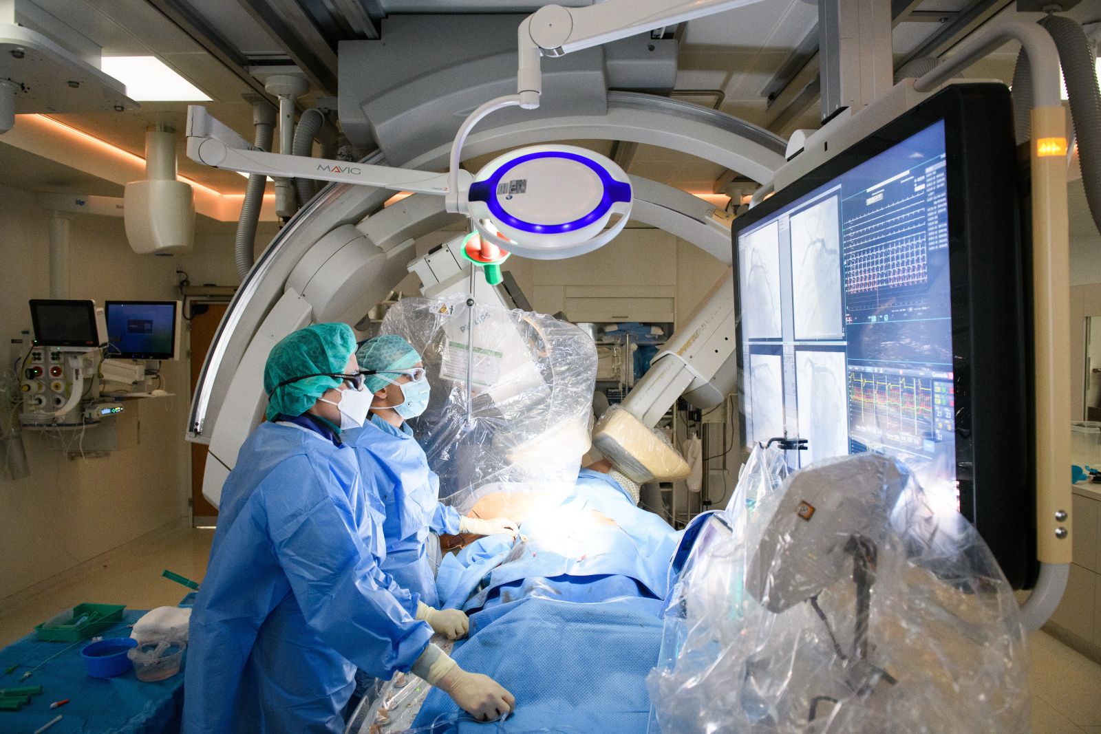
Specialists diagnose and treat heart diseases in around 3,000 patients in the cardiac catheterization laboratories at the University Hospital Zurich (USZ) every year. A look over the shoulder of cardiologist Barbara Stähli into her patient’s heart.
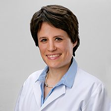
“Good Morning Ms. Furrer,” says Barbara Stähli to greet her patient. The cardiologist and deputy head of the cardiac catheterization laboratories at the USZ is specialized in complex coronary interventions, functional coronary diagnostics, i.e. the examination of the function of the small and large coronary vessels, and the replacement of heart valves by means of catheter interventions. Daniela Furrer*, her first patient this morning, undergoes functional coronary diagnostics.
Further medical examination at the University Hospital Zurich
Ms. Furrer has had heart problems for a long time. She is suffering from a constant feeling of pressure, and she has difficulty breathing. A stabbing pain occurs sporadically. Initial examinations by her doctor had not revealed any abnormalities. Now, the cause of her symptoms is to be identified in the cardiac catheterization laboratories at the USZ. “We examine Ms. Furrer’s coronary vessels,” Barbara Stähli explains. “We distinguish between large and small coronary vessels. Both supply the heart with oxygen. If the smallest coronary vessels are tightened temporarily or narrowed, we call it a microvascular dysfunction. Although Ms. Furrer’s symptoms are typical for such a dysfunction, we are now examining the cause in detail in order to be able to treat the symptoms effectively.”
Minimally invasive examination
Ms. Furrer’s examination is done in the cardiac catheterization laboratory, which looks similar to an operating room. The risks of cardiac catheter examinations and interventions are low. More than 3,000 patients are examined and treated by specialized cardiologists in the cardiac catheterization laboratories at the USZ every year. A cardiac catheter examination is a minimally invasive examination of the heart that can be done to diagnose a variety of diseases and, in certain cases, to treat them in the same session. For this purpose, a thin, flexible plastic tube, i.e. the heart catheter, is pushed through a blood vessel to the heart. The catheter is usually inserted via a vessel in the groin or in the wrist. Contrast media can be injected through the catheter into the coronary vessels that supply blood to the heart in order to make the vessels visible and to examine whether they are narrowed. The X-ray images are directly assessed via an X-ray machine, allowing the position of the catheter to be constantly monitored. A cardiac catheter examination is the result of the team effort of specialists. During the examination, further specialists provide the required materials in a sterile manner, administer medication, monitor blood pressure and heart rate or record the entire procedure.
The team is ready 24 hours a day
The most common reason for a cardiac catheter examination is symptoms or evidence of a circulatory disorder of the heart muscle in a specific, non-invasive test. If one or more vessels are narrowed, they can be expanded with the help of a balloon inserted via the catheter and kept open with a stent. An acute vascular occlusion is a life-threatening heart attack. Specialists working in the cardiac catheter laboratories at the USZ are ready 24 hours a day to treat patients suffering from a heart attack. This makes it possible to reopen an occluded coronary vessel as quickly as possible. Other specific catheters are used to measure a pressure drop across a constriction in a coronary vessel or to generate high-resolution cross-sectional images of the coronary vessels, which allows precise planning and execution of interventions.
Ms. Furrer is a little nervous
Daniela Furrer was already prepared for the examination in the anteroom. She is a little nervous as she knows that a small tube will be pushed into her heart. This is why she received a mild sedative. The procedure itself is painless. She will hardly feel any of it, and only the entry point for the catheter will be anaesthetized. Like all patients, she has a venous access so that medication could be administered quickly if needed. Ms. Furrer is taken to the cardiac catheter laboratories, where her blood pressure and heart rate are continuously monitored and the oxygen level in her blood is checked using a finger clip. Then the examination begins.
Functional diagnostics allows clarity
There are no abnormalities in the large coronary vessels. Barbara Stähli therefore examines the function of Ms. Furrer’s small coronary vessels. They show a tendency to tighten temporarily. She suffers from what is known as coronary microvascular dysfunction. The reason for her symptoms is clear now. “We carry out this special examination of the smallest vessels at the USZ when there is clinical suspicion of a coronary vessel dysfunction,” Barbara Stähli explains. “It is important to detect coronary vessel dysfunction in order to avoid unnecessary further examination and to initiate the right treatment. The patients – around 60 to 70 percent of those affected are women – often suffer for a long time before the disease is detected.” Ms. Furrer is happy to have had the examination, which took around 15 minutes and was, in fact, painless. She can go home the same day. Her microvascular dysfunction can be easily treated with medication. Her symptoms will probably subside or disappear in a short time.
* Fictitious name