With the help of computed tomography (CT) and magnetic resonance imaging (MRI) examinations, it is possible to depict almost all aspects of the anatomy and function of congenital diseases of the heart and the great vessels. The strength of CT examinations is the rapid and detailed visualization of the anatomical structures of the heart as well as the coronary vessels and the large thoracic and abdominal vessels(Figure 1).
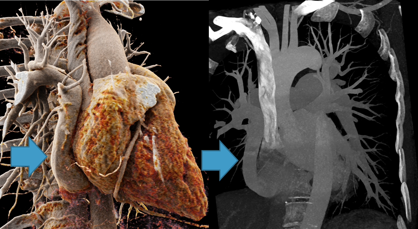
Figure 1: CT angiography of a patient with partial pulmonary vein malformation. The venous drainage of the right upper lobe of the lung and the middle lobe incorrectly flows into the inferior vena cava (blue arrow), corresponding to a so-called “scimitar vein”.
Cardiac MRI can also be used to assess the function of the heart chambers and the tissue properties of the heart muscle. MRI is the reference standard, particularly for quantifying the size and ejection fraction of the ventricles, and thus provides the basis for accurate progression assessments(Figure 2). Flow measurements can be used to investigate diseases of the heart valves and to detect faulty connections (shunts) between vessels or heart chambers and determine the severity(Figure 3). Cardiac MRI does not require ionizing radiation and is therefore particularly suitable in cases where regular follow-up checks are required.
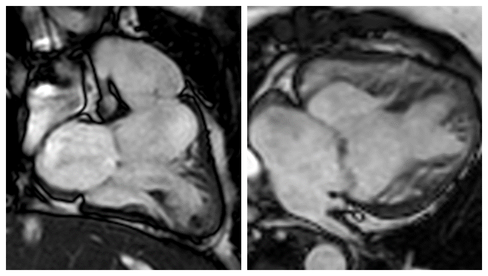
Figure 2: MRI functional images (SSFP cine) of a patient with hypoplastic left heart syndrome and surgical conversion of the supply of the systemic circulation to the right ventricle (“Fontan circulation”).
Patients with “metal in the body” (such as electrodes, pacemakers and valve prostheses) can also receive a cardiac CT scan and, in most cases, a cardiac MRI scan without any problems.
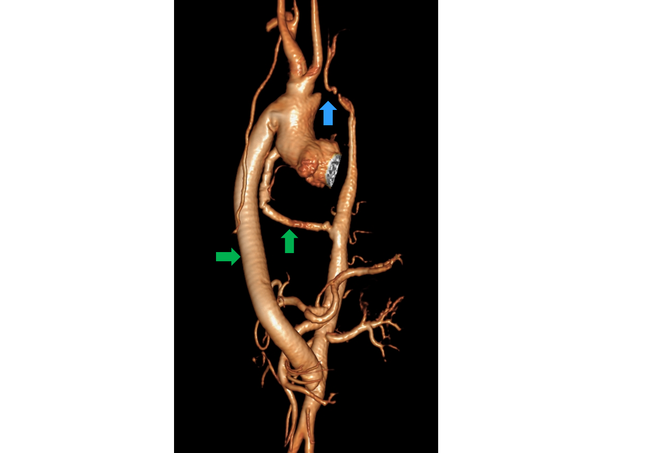
Figure 3: MRI angiography of the aorta of a patient with severe aortic isthmus stenosis (blue arrow) and surgical placement of two extra-anatomical aortic bypasses (green arrows).