Overview
Significant progress has also been made in reducing radiation doses. Using state-of-the-art equipment, the coronary arteries can be imaged with a dose of less than 1 mSv.
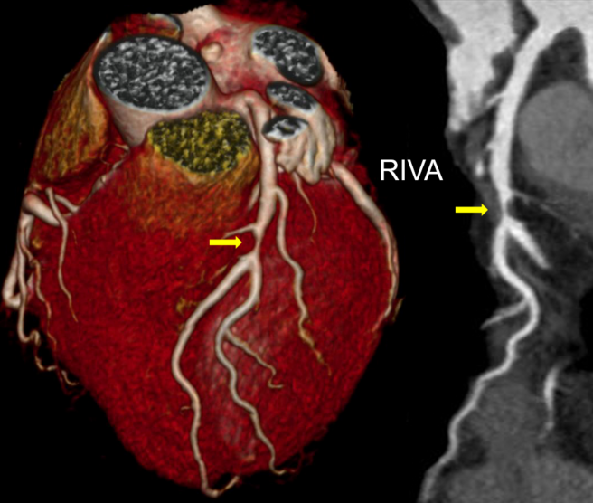
Even dynamic CT angiography of the heart—such as that required to assess the success of interventional closure of the left atrial appendage—or CT examinations following interventional mitral valve repair can now be performed with a very low radiation dose.
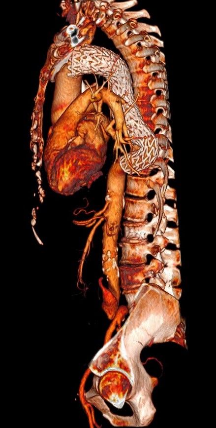 Last year, clinical research focused on so-called “kidney-friendly” CT angiography, which uses only small amounts of iodine-containing contrast agents. This method takes advantage of the specific attenuation behavior of iodine as a function of the energy of the X-rays.
For example, today, only about 20 ml of contrast agent is administered for a CT pulmonary angiography to diagnose pulmonary embolism, and an ECG-triggered CT angiography of the aorta for planning a TAVI (transcatheter aortic valve implantation) or an endovascular procedure is now performed using only 30 ml of contrast agent.
Given the high prevalence of renal insufficiency among patients eligible for TAVI and endoaortic procedures, this reduction in the amount of contrast agent is a major advantage.
An accurate pre-procedural assessment of the anatomy is essential for planning TAVI procedures. CT allows for highly accurate measurement of the aortic annulus and the distance between the coronary ostia and the annular plane, which is crucial for selecting the correct prosthesis size. In addition, CT allows for accurate visualization and quantification of the diameter of the pelvic vessels to help plan the access route (femoral vs. transapical).