Digital planning, customized implants and high-precision surgery help to restore function and appearance in cases of severe facial injuries.
Nerves, muscles, tendons and blood vessels with important functions lie close together in the skull and especially in the face. These structures usually play alongside each other and thus enable breathing, chewing, speaking, seeing and facial expressions. An accident or illness that leads to functional limitations in this area is therefore serious. If there are also visible consequences, this is doubly stressful for patients. “In reconstructive procedures following facial injuries, the restoration of function is the primary criterion,” says Maximilian Wagner, Head Physician and Deputy Director of the Clinic for Oral and Maxillofacial Surgery at the USZ. “At the same time, we always strive to achieve the best possible aesthetic result for our patients.” The clinic treats patients when bone structures are injured or large sections of soft tissue and bone need to be replaced. On a case-by-case basis, cooperation also takes place with specialists from the Eye Clinic, Ear, Nose, Throat and Facial Surgery, Plastic Surgery and Neurosurgery.
The facial skull – a complex structure
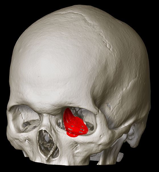
Fractures of the facial skull are among the most common injuries treated surgically in the clinic. “The bone of the eye socket is particularly thin,” explains Maximilian Wagner. “That’s why it breaks quite quickly and often in several places at once. And because there are naturally very delicate bony structures at this point, these fractures do not heal well or not in the optimum position. This can have dramatic consequences for vision if the entire eye is no longer in the correct position. The patient then constantly sees double vision, for example. ” An orbital fracture, as this injury is called in technical jargon, must therefore be treated surgically in many cases. Another reason for reconstructions are bone and soft tissue defects caused by diseases. Cancers in the facial area can lead to the loss of bone, soft tissue or both. For the safe removal of tumors, in some cases a lot of substance has to be removed – with corresponding consequences for the appearance and functional limitations.
Implants and artificial
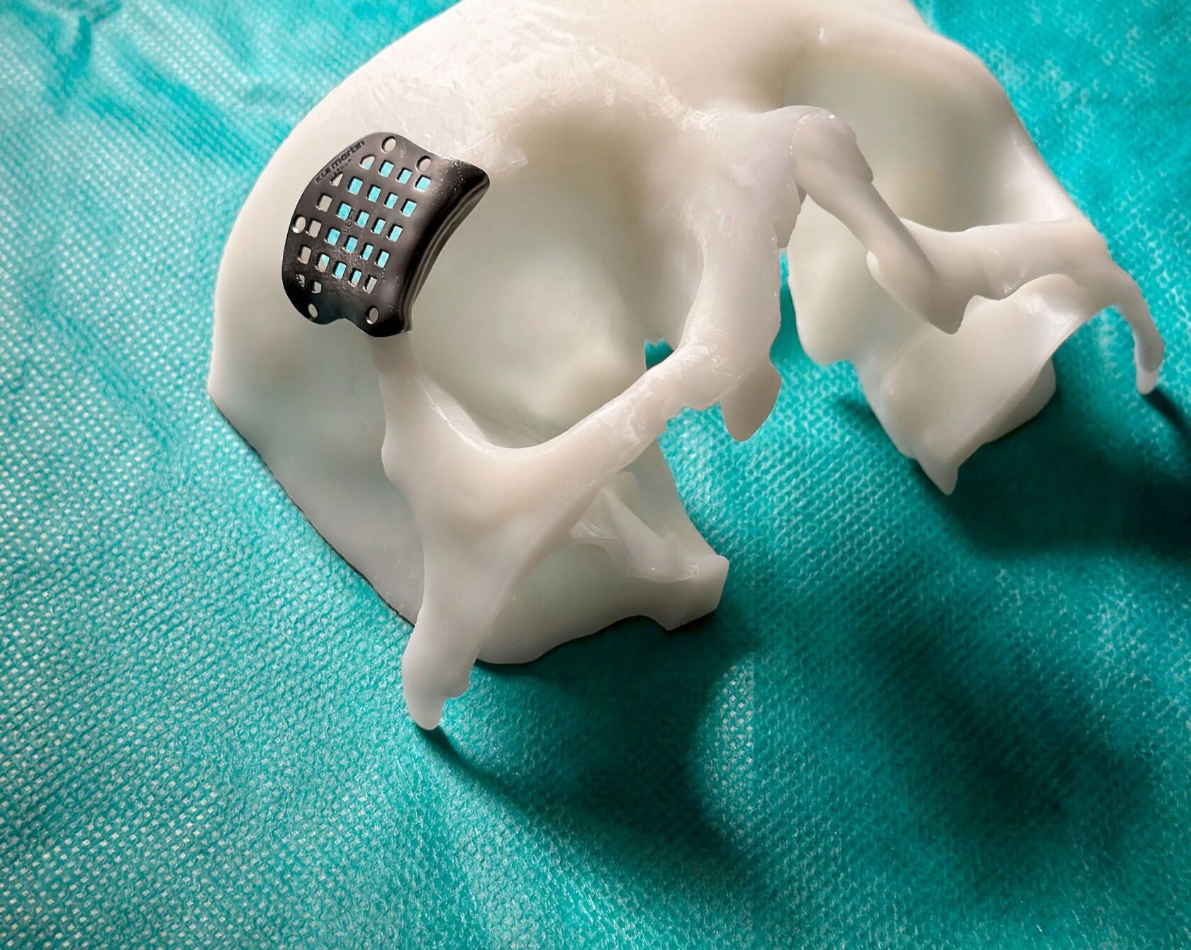
Tissue as a substitute Various methods are used for reconstruction. Artificial or autologous tissue can be used to reconstruct defects. Missing bony structures can be reconstructed using titanium implants or by transferring the patient’s own bone from the skull, pelvis or fibula. ” The possibilities for restoration have made enormous progress,” says Maximilian Wagner. One reason for this is new tools in the planning and preparation of operations. Surgeons have been able to use digital planning tools for this for around nine years. Implants were already being used to replace destroyed bone structures. However, the ones used today are manufactured with high precision and fit the patient perfectly. The basis for this is modern imaging.
The implants are ready after a short time
The data for the implants is transmitted to the manufacturer, where the elements are sintered from titanium, for example, with high precision using a laser. After a few days they arrive at the USZ. “This waiting time, for example after an accident, is not a disadvantage,” explains Maximilian Wagner. “Swelling usually persists for a few days. Unless an operation is absolutely necessary in an emergency, we therefore deliberately wait some time until the swelling has subsided and is not distorting the image.” In order to test the exact placement and accuracy of fit of the implants in advance or to manufacture them on site, a three-dimensional model of a patient’s jaw, for example, is also created from plastic using the image data if necessary, on which the implant can be placed one-to-one before the operation. Because the data collected once can be used for the entire planning process, there is no need for repeated and radiation-intensive examinations. Another advantage is that if the teeth are also affected by the injury, subsequent dental treatment can be planned and prepared in consultation with the treating dentist. This shortens the duration of treatment for these patients by many months.
The risk of complications decreases enormously
This time-consuming process is worthwhile. “We clearly achieve better results for our patients,” explains Maximilian Wagner. “The operations take less time, we rarely experience unforeseen events during the operation and, thanks to the high level of precision, we have been able to greatly reduce the risk of post-operative visual impairment and other complications, for example.” In order to achieve the highest possible precision, the surgeons compare the placement of the implant with the planning data during the operation using three-dimensional imaging.
The images show the patient the result in advance
With this process and its experience, the Department of Oral and Maxillofacial Surgery is a world leader in the restoration and reconstruction of bony structures with patient-specific implants. This means that not only accident patients from a wide area are brought directly to the USZ. Some of the patients also come for advice on a second operation or to have it performed here. “There are various reasons why, for example, an injury could not be optimally treated and a fracture therefore healed in a malposition,” explains Maximilian Wagner. “The result can then perhaps be corrected and improved with a planned procedure.”
The reconstructions are planned virtually.
Visible memories can be a burden
The patient decides what is done. Especially with aesthetic adjustments, what bothers someone or not is very different. “Being reminded of an accident or illness every time you look in the mirror, constantly feeling curious glances, can be very stressful, even if function is not impaired.” Simulating the result in advance is also helpful for these patients. This gives you a good basis for deciding whether the improvement that can be achieved with further surgery meets your wishes and expectations.
Operations remain challenging
“Reconstruction of the eye socket is a very demanding surgical procedure because the area around the eye is very delicate,” says Wagner. “We generally want to avoid visible scars, which is why we insert the implants on the inside of the lower eyelid. In the case of jaw injuries, the upper and lower jaws must be aligned in order to close properly. Reconstructions in which a lot of tissue and bone has to be replaced on both sides and we have to rebuild both are also difficult. We want the best result for every patient. However, we are always particularly pleased when we succeed in achieving this in particularly difficult initial situations.”