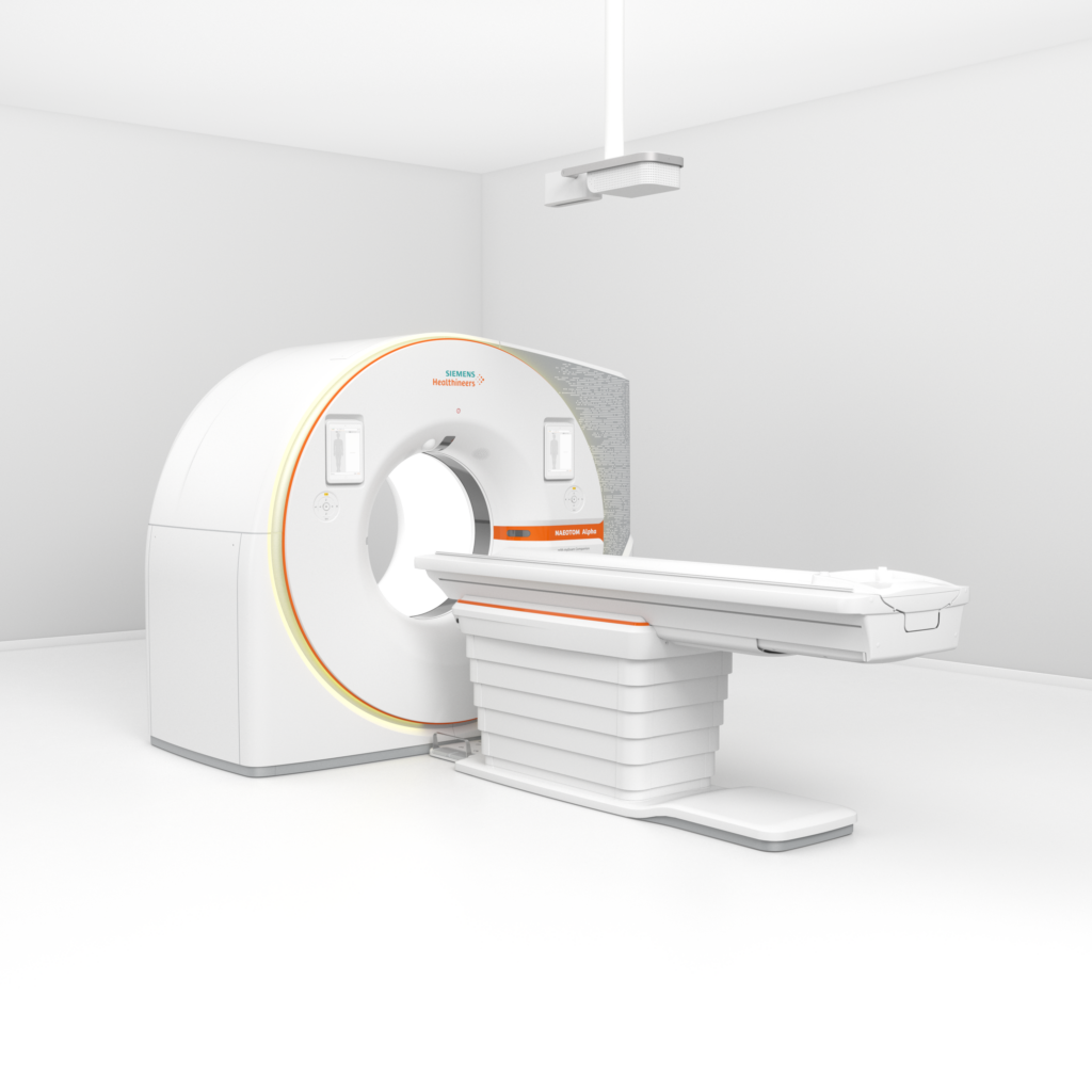
Sharp images like never before thanks to a tiny single crystal. The first of three CT scanners worldwide with new photon-counting technology is located at the University Hospital Zurich (USZ). Patients benefit from improved diagnostics and less stressful examinations, while the manufacturer benefits from the experience of the practice specialists at USZ.
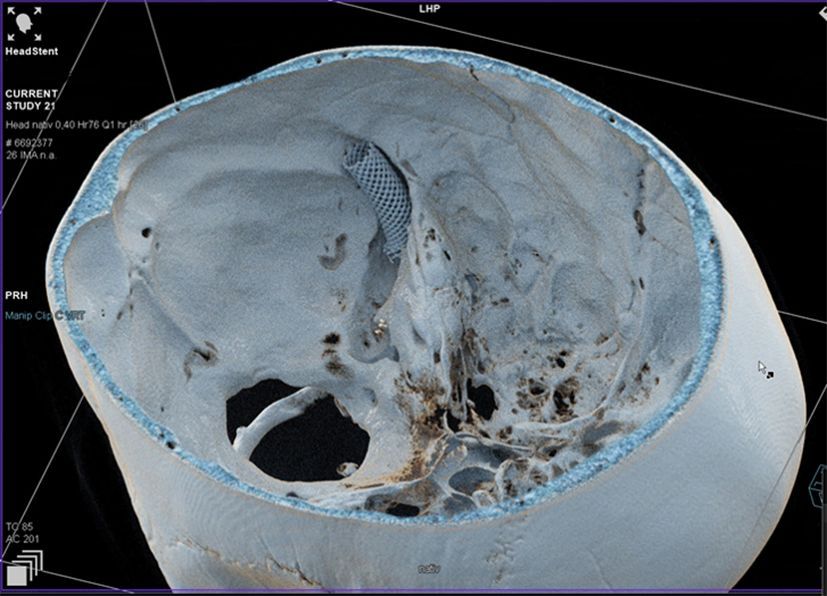
From the outside, the new CT scanner looks to laypersons just like the well-known, almost room-filling CT scanners which can be used to take three-dimensional images of body structures. The revolution only becomes visible when looking at the images of the new CT: pin-sharp, ultra-fine structures in bones or tissue that were previously only blurred or not visible at all.
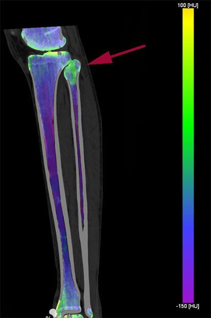
This is made possible by a tiny single-crystal cadmium telluride (CdTe) that is only the size of a pinhead in the device. The crystal cultivated in a laboratory has the ability to convert individual X-rays directly into electrical pulses. In a two-stage process, conventional CT scanners first convert the X-rays into visible light which is detected by a light sensor and only then generates the image. As a result of this intermediate step, important information about the energy of the X-rays is lost. As a result, the contrast is reduced and the images are less sharp. The new detector converts X-ray photons directly into fully digital electrical signals which are then counted without the loss of any information. This technique, known as “photon counting,” significantly improves contrast and image sharpness – a similar effect to the improved image resolution of digital photos with a high number of pixels.
Experience from practice and research provides valuable information for improvement
The first of a total of three pilot production units in the world with this new technology had been in clinical use at USZ since the spring of 2021, before it was officially presented to the industry on November 15. USZ has been collaborating with Siemens Healthineers, the manufacturer of the device, on research and development for many years. “The experience gained and requirements identified during clinical operation are essential for improving medical devices. Our critical feedback and systematic and scientific tests are thus incorporated into further development at an early stage,” says Hatem Alkadhi, Senior Consultant at the Department of Diagnostic and Interventional Radiology at USZ, describing the cooperation between manufacturers and specialists with practical experience. The test of a pilot production unit is at the end of the development phase. The devices are approved and ready for production. The aim of this phase is for specialists to continue using the new device in day-to-day clinical practice for an extended period of time and to gain practical experience with it that may be helpful to other new users. Hatem Alkadhi and his team of radiologists and medical technical radiology assistants have already been able to test several such pre-production models. They are accordingly experienced in recognizing and describing problems – and often also making suggestions for improvement at the same time, for example for adjustments to operation and the work process. Research results are also continuously shared with the physicists at Siemens Healthineers, leading to improvements in image quality, for example. “At the same time,” says Alkadhi, “our patients can benefit from the most advanced technologies long before they are launched on the market. In addition, the devices are already available to us for research at this stage.” Access to state-of-the-art technology is also a valuable asset for the training of young doctors.
More precise images help to achieve targeted treatment
The “Super CT” has proven its worth in practice: “With this technology, we can see for the first time fine structures in organs, tiny auditory ossicles and hairline cracks in bones that were previously hidden to us or could only be spotted out of focus because the technology was reaching its limits.” Hatem Alkadhi is convinced that this dramatically improved imaging will have a direct effect on treatment. The use of photon counting in the Medical Department is more than just a technical gimmick. “The more precise the diagnosis, the more accurate and targeted doctors can tailor the therapy. Imaging plays a key role in this,” says Alkadhi. Sharper images mean, for example, that tumor types can already be identified by the CT, or that patients with a heart condition are spared the examination with a cardiac catheter because the CT alone provides the necessary information. Follow-up examinations of vascular and organ diseases are therefore more informative. “This is a massive advance, especially in the case of lung diseases, because the photon-counting CT makes it even easier to image lung tissue.” However, Hatem Alkadhi sees further advantages for patients: “In all examinations, we can extract so-called multi-energy information from the images. This means that we receive additional information about the composition of tissue and pathological processes that can be used for the benefit of diagnosis and therapy. In addition, there is another important effect for patients: in some cases, the radiation dose for the examinations can be reduced by up to 40%, and the use of contrast agents can also be significantly reduced. This is an important and significant improvement for people with kidney problems or younger patients.”