Operations to remove tumours in the throat or thoracic cavity sometimes entail cutting through major lymphatic vessels. Until now, these vessels could not be restored, and the consequences were potentially fatal. A team working with plastic surgeon Nicole Lindenblatt of the USZ has, for the first time in Switzerland, reconstructed the largest lymphatic vessel in the human body.
The thoracic duct is the largest lymphatic vessel in the human body. The thoracic duct travels from the lumbar region to the throat by way of the thorax. During tumour operations in the throat or thoracic region, it is vulnerable to injury or separation. The result is a heavy secretion of up to two litres of milky lymphatic fluid per day from the surgical wound. And it is all but impossible to stop the flow. The fluid accumulates in the wound or thorax, with potentially fatal consequences for patients. Prof. Dr. med. Nicole Lindenblatt, senior physician in the Department of Plastic Surgery and Hand Surgery and Deputy Director of the hospital, has now succeeded in restoring the thoracic duct. Following an operation to remove a throat tumour, she connected the patient’s lymphatic duct under a microscope to his veins, thereby ensuring proper drainage of the lymph.
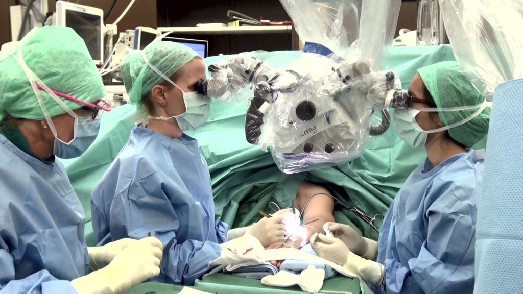
Super-microsurgery: needles and thread finer than a human hair
For this procedure, the thoracic duct is visualised with the use of a blue and a green, fluorescent dye, which are injected through the skin into the patient’s arms or legs or administered to the patient through a stomach tube. The dye is absorbed into the lymph, rendering the leak visible under the microscope. The openings in the lymphatic vessels are then connected super-microsurgically to a neighbouring vein to restore physiological drainage.
This can be done only thanks to the new technological capabilities of super-microsurgery. An example of the new technology in use is a specialised fluorescence microscope with up to 50x magnification. And the other instruments that are used are super-fine as well. The thread that is used to sew the vessels together is thinner than a human hair. The needle is two millimetres long and has a diameter of 0.05 millimetres. “From a surgical standpoint, it has always been considered virtually impossible to repair the thoracic duct, as it is sometimes only one millimetre in diameter and impossible to find in the wound or scar,” explains Nicole Lindenblatt. The usual procedure until now has been to attempt, often unsuccessfully, to ligate the lymphatic vessel. “This operation requires super-microsurgical skills,” adds Nicole Lindenblatt.
Elsewhere in the world the procedure has been performed only a few times, but never before in Switzerland. The procedure can help not only patients who must undergo surgery for tumours, cardiac disorders, or trauma, but also patients with congenital malformations of the thoracic duct.