White skin cancer is around ten times more common in Switzerland than malignant melanoma. Early detection remains key, as the chances of recovery are excellent if diagnosed in time. New imaging techniques such as AI-supported LC OCT and proven Mohs surgery enable precise, safe and tissue-sparing diagnostics and treatment at the USZ. They help to avoid unnecessary interventions and improve the care of high-risk patients in the long term.
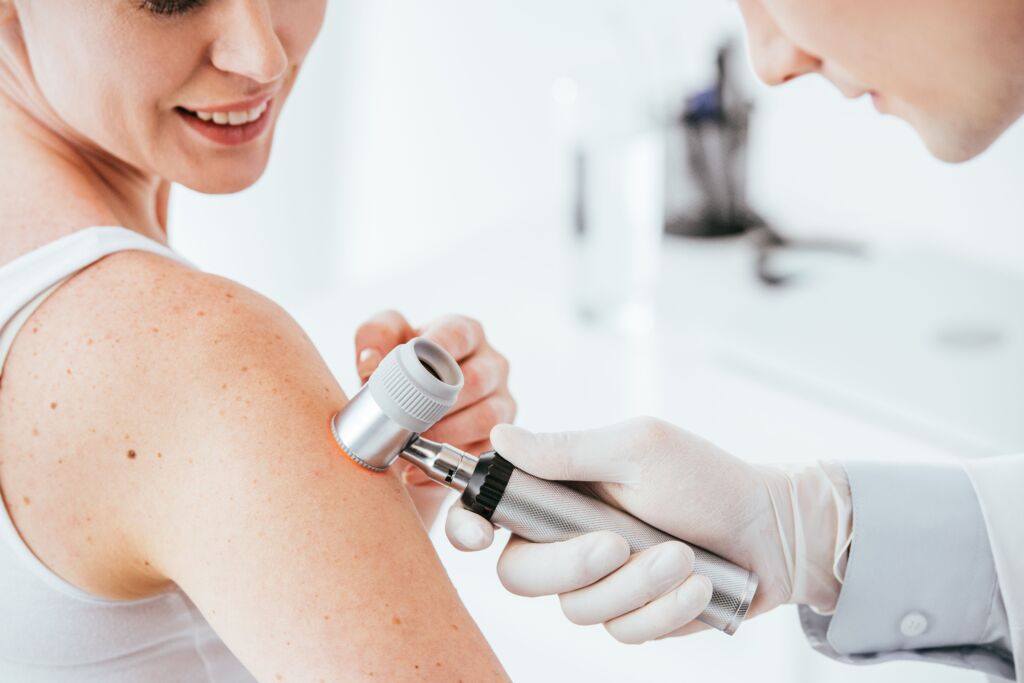
White skin cancer is one of the most common forms of cancer. If it is detected early, the chances of recovery are very high. Preventive skin inspections are therefore advisable for people at risk. Suspicious skin changes should also always be assessed by a dermatologist. In addition to a careful clinical examination, modern imaging techniques are now available which improve the diagnosis without the need for an immediate biopsy.Diagnostics with AI in dermatology at the USZ
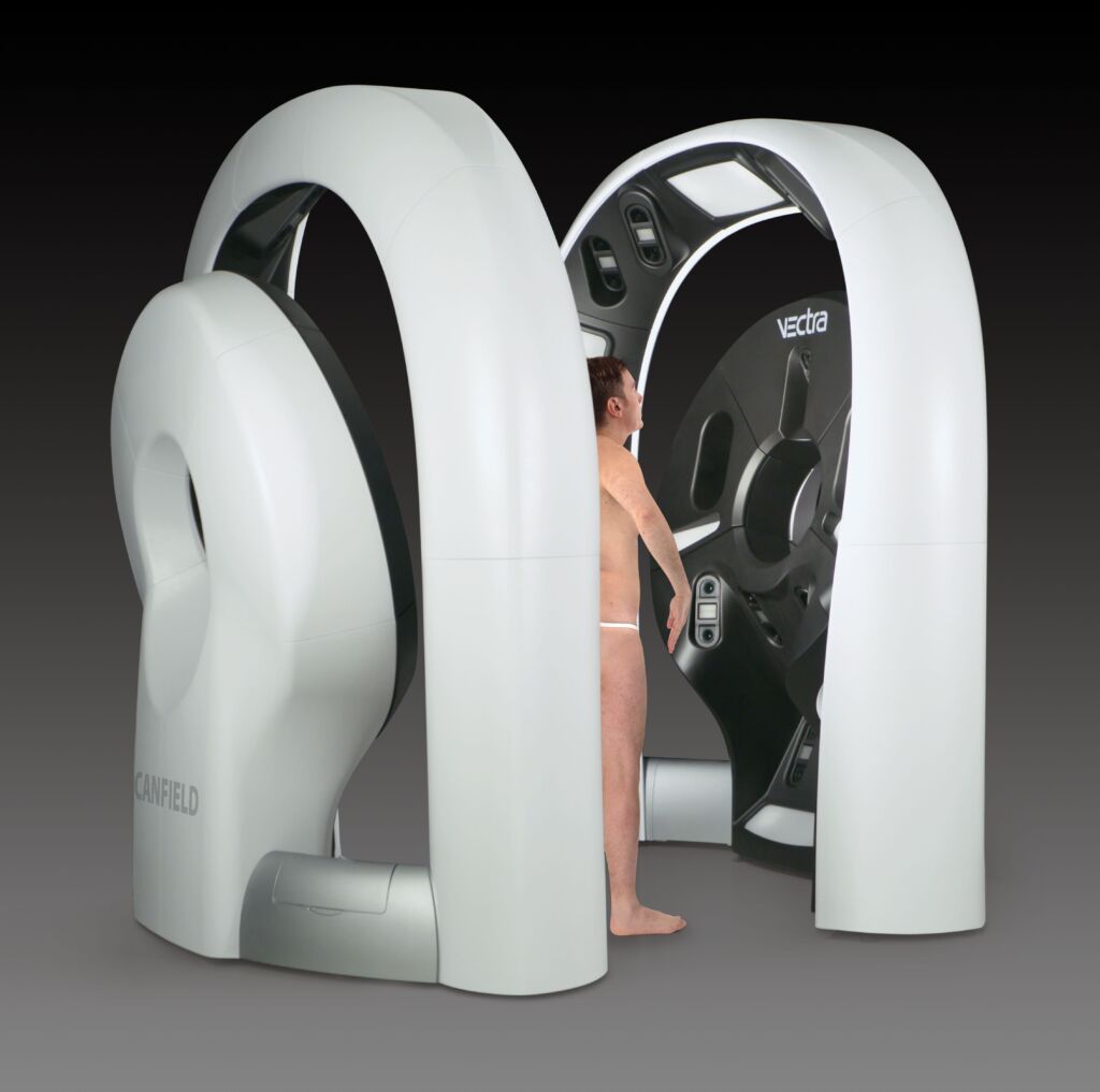
Line-field confocal optical coherence tomography (LC-OCT), a modern, non-invasive imaging technique, has recently been introduced at the Dermatology Clinic of the USZ. Similar to histology, it provides high-resolution, real-time images of the skin structure at the cellular level. This CE-certified and AI-supported technology supports doctors in the early and reliable diagnosis of white skin cancer, namely basal cell carcinomas and spinaliomas, as well as precursors, actinic keratoses, without the need for a surgical biopsy.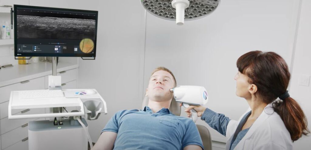 In addition, the method enables preoperative monitoring of tumor margins, allowing surgical interventions to be planned and performed in a more targeted manner. It also makes it possible to monitor the progress of therapies in real time and assess their success immediately.
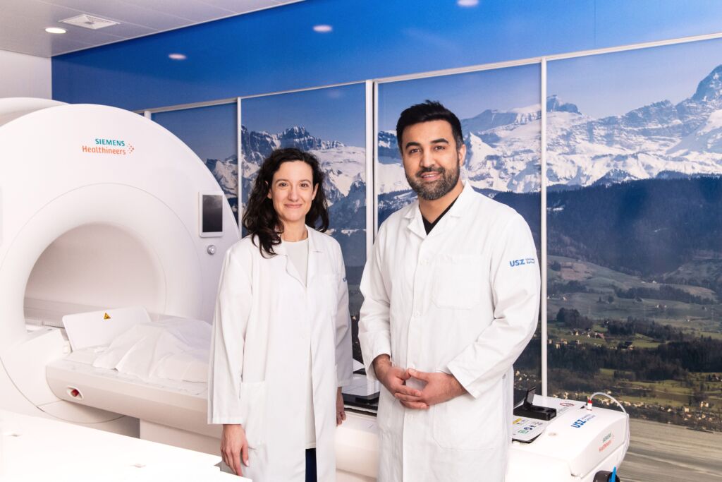
"Line-field confocal optical coherence tomography (LC-OCT) represents a significant advance in non-invasive imaging of white skin cancer and can significantly improve patient care and reduce unnecessary interventions." Dr. Christian Greis, Senior Attending Physiciant and Head of Dermatosurgery, Dermatology Clinic
Mohs surgery is the gold standard
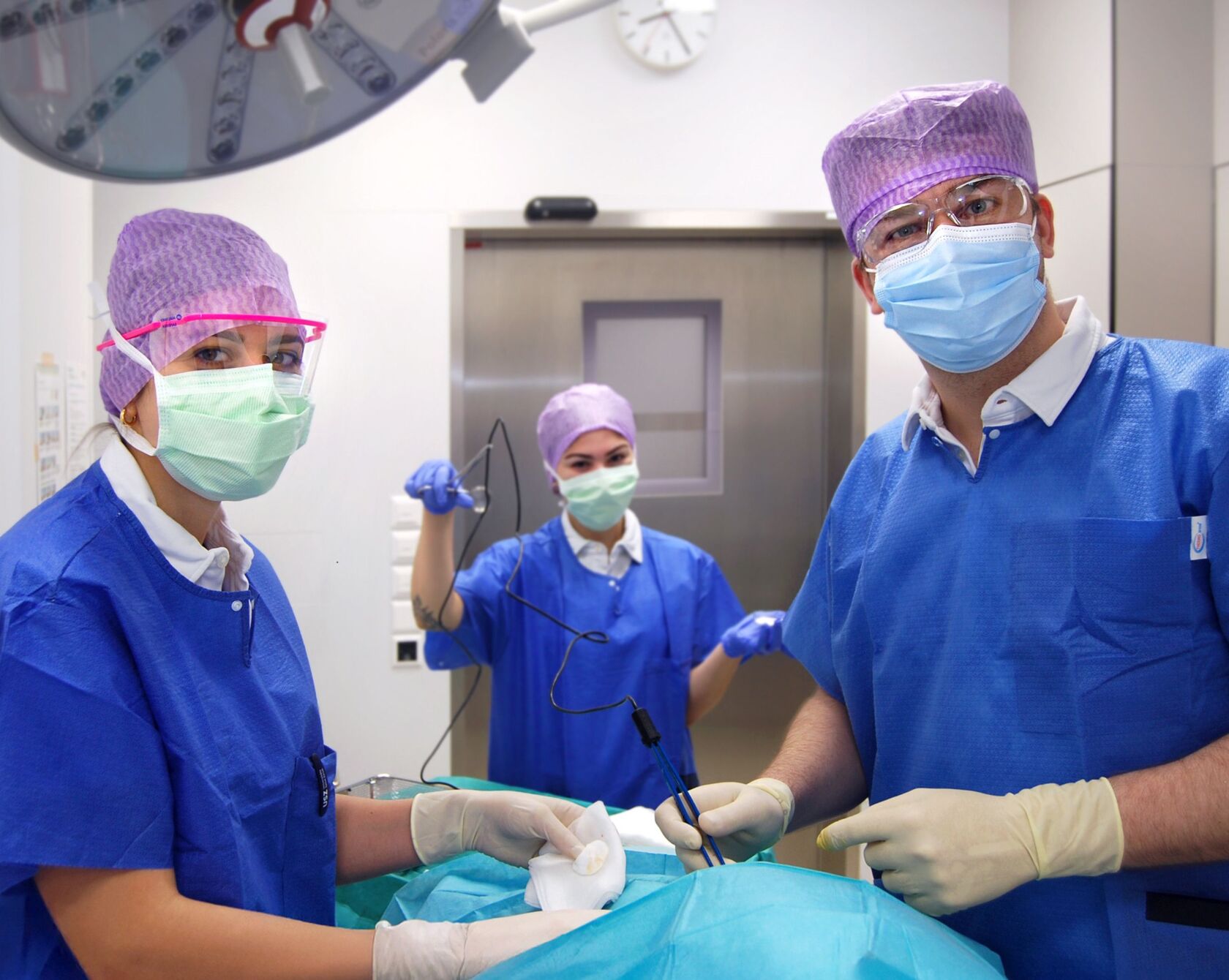
The Mohs technique has been used successfully at the USZ for years. The "Tübingen cake" (incision margin control in formalin and kerosene) was already introduced in the dermatology clinic in the second half of the 1990s and continues to be used primarily for incision margin control in large tumors or tumors outside the central facial region. Since the introduction of the rapid incision technique in 2005, over 15,000 patients have been successfully operated on using micrographically controlled surgery according to Dr. F. Mohs. Thanks to the complete microscopic assessment of the incision margins, it is particularly indicated for- Tumors on the face (especially eyelids, nose, ears, lips)
- Recurrent tumors or clinically indistinct tumors
- Histologically aggressive subtypes (e.g. sclerodermiform basal cell carcinoma)
"The ability to produce dermatopathological findings directly in-house strengthens our diagnostic reliability, speeds up decision-making processes and improves patient care in the long term." Prof. Jürg Hafner, Deputy Clinic Director and Head of Dermatosurgery, Dermatology ClinicThe surgical team treats around one in three patients with basal cell carcinoma or spinalioma using Mohs surgery in one of three operating theaters at USZ Airport. Patients with advanced white skin cancer or the very elderly are operated on once a week at the USZ Campus in an inpatient setting. We work closely with internal specialists, such as plastic surgeons, ophthalmologists and ear, nose and throat specialists, and not only at the regular tumor boards. In the case of white skin cancer, Mohs surgery massively reduces the risk of lengthy second and third operations compared to standard surgery. This saves patients a lot of suffering and saves you as a GP time in follow-up care.