LC-OCT: Precise skin diagnostics with AI and without biopsy

LC-OCT (Line-field Confocal Optical Coherence Tomography) is a non-invasive, AI-supported imaging technique that provides vertical, horizontal and three-dimensional real-time images of the skin. At the USZ, the method is used in particular to diagnose white skin cancer. The clinical evidence is currently strongest for basal cell carcinomas.

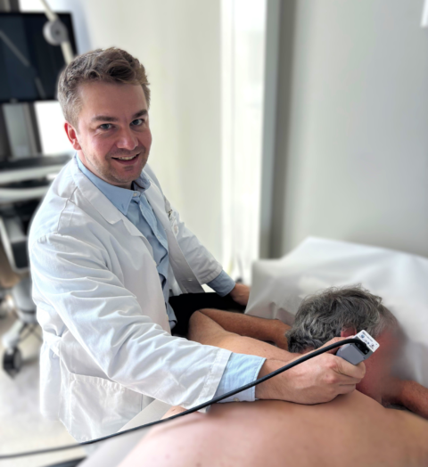
Line-field Confocal Optical Coherence Tomography (LC-OCT) is a modern, non-invasive imaging technique available at the Dermatology Clinic of the University Hospital Zurich. The method enables high-resolution real-time images of the skin and can help to assess suspicious skin changes more precisely – in selected situations without immediate tissue removal.

LC-OCT is currently particularly relevant for white skin cancer, especially basal cell carcinoma. This is where the scientific data is strongest, and this is also where AI-supported image analysis is at its most advanced: it supports the medical assessment of unclear lesions, increases diagnostic certainty and can standardize clinical decision-making.

Arzt mit Patient bei LC-OCT: Präzise Hautdiagnostik

What is LC-OCT?

LC-OCT combines the principles of optical coherence tomography and confocal imaging. The procedure generates histology-like images in vivo and in real time. Vertical sections, horizontal planes and three-dimensional volumes of the epidermis and the superficial to middle dermis area can be visualized [5,6].

What does AI do for basal cell carcinoma?

The AI analyzes vertical LC-OCT images for BCC-typical patterns during the examination. It outputs a probability value and optionally a color code that marks the areas of the image that appear suspicious.

When can LC-OCT with AI be useful?

  • for clinically or dermatoscopically unclear lesions with suspected basal cell carcinoma
  • if an additional, standardized second assessment of the LC-OCT images is helpful
  • for blurred skin changes, especially in the facial area
  • for preoperative assessment of the horizontal extent of the tumor
  • for therapy planning when weighing up between conservative and surgical treatment
  • for follow-up after surgery, radiotherapy or systemic therapy, if scar tissue and residual or recurrent tumor are difficult to distinguish clinically

Your advantages

  • Non-invasive and generally painless examination
  • histology-like real-time images directly in the consultation
  • AI-supported assessment especially for lesions suspected of being BCC
  • Greater diagnostic certainty in unclear cases of basal cell carcinoma
  • Support for treatment decisions and preoperative planning
  • Potentially fewer unnecessary diagnostic biopsies with high clinical safety

What are the limits of LC-OCT?

LC-OCT is a very helpful additional tool, but does not always replace histology. This also applies to AI assistance. In the case of deep-seated, high-risk or still unclear findings, the biopsy or histological examination remains the reference standard.

Frequently asked questions

LC-OCT provides high-resolution real-time images of the skin. The AI additionally analyzes some of these images for BCC-typical patterns and supports the physician’s interpretation. A biopsy, on the other hand, removes tissue for histopathological examination. LC-OCT and AI can supplement or avoid a biopsy in selected cases, but do not replace it in principle.

No. The examination is usually painless and is performed without anesthesia, incision or tissue removal.

Yes, in selected cases. LC-OCT can help to better estimate the horizontal extent of blurred basal cell carcinomas in particular and make surgical planning more precise. Initial work also suggests that AI-supported localization and edge assessment could provide additional support in the future.

Yes. Initial studies and clinical experience suggest that LC-OCT can be helpful in monitoring progress, for example when it is clinically unclear whether scar tissue, therapy sequelae or residual tumor is present.

Literature

  1. Gust C, et al. Line-Field Confocal Optical Coherence Tomography Increases the Diagnostic Accuracy and Confidence for Basal Cell Carcinoma in Equivocal Lesions: A Prospective Study. Cancers (Basel). 2022;14:1082.
  2. Ruini C, Schuh S, Pellacani G, et al. Line-field optical coherence tomography: in vivo diagnosis of basal cell carcinoma subtypes compared with histopathology. J Biophotonics. 2021;14:e202000449.
  3. Paradisi A, et al. Preoperative evaluation of high-risk basal cell carcinoma with line-field confocal optical coherence tomography (LC-OCT) reduces Mohs micrographic surgery stage number: A case-control study. EJC Skin Cancer. 2024;2:100015.
  4. Dubois A, Levecq O, Azimani H, et al. Line-field confocal optical coherence tomography for high-resolution noninvasive imaging of skin tumors. J Biomed Opt. 2018;23:106007.

Responsible professionals

Christian Greis, MBA, Dr. med.

Senior Attending Physician, Department of Dermatology

Tel. +41 44 255 11 11
Specialties: Dermatosurgery (Mohs surgery), Laser medicine & aesthetics , Telemedicine & artificial intelligence (digital dermatology)

Fabienne Fröhlich, Dr. med.

Attending Physician, Department of Dermatology

Tel. +41 44 255 11 11
Specialties: Dermatosurgery (Mohs surgery), LC-OCT

For patients

If you have any questions or would like to make an appointment, please contact the Dermatology Clinic of the USZ. The central telephone number of the USZ is +41 44 255 11 11.

For referrering physicians

Department of Dermatology
Raemistrasse 100
8091 Zurich

Tel. +41 44 255 11 11

Responsible Department