Overview: What is skin cancer?
In skin cancer, cells in the skin begin to grow uncontrollably. Depending on which cells are involved, a distinction is made between different forms of skin cancer. The most important are:
Basal cell carcinoma and spinal cell carcinoma are collectively referred to as white skin cancer.
Skin cancer – frequency and age
Black skin cancer is the fifth most common type of cancer in Switzerland and accounts for seven percent of all cancers. Every year, around 2,800 people in Germany contract the disease. Almost a quarter of them are younger than 50 when the malignant tumor is discovered.
White skin cancer is significantly more common than malignant melanoma, even if the exact number of new cases in Switzerland each year is not known – most cancer registers do not record cases of white skin cancer. However, experts estimate that 20,000 to 25,000 people in Switzerland are diagnosed with some form of white skin cancer – usually basal cell carcinoma, less frequently spinal cell carcinoma – every year.
The average age of those affected by basal cell carcinoma is 60, although an increasing number of younger people are also developing the disease. Men and women are affected about equally often. In contrast, spinal cell carcinoma occurs more frequently in men than in women. On average, those affected are 70 years old at the time of diagnosis.
Online skin check: Quick and convenient from home
Send a photo of the affected skin area and fill out the short questionnaire. Within 24 hours on weekdays, you will receive a reliable diagnosis from our experts. Your data will be transmitted to us encrypted and treated confidentially.
Skin cancer: causes and risk factors
The most important trigger for all forms of skin cancer are UV rays (in sunlight or solarium). They damage the genetic material (DNA) in the cells. If the body is unable to repair this damage, the affected cell can mutate into a cancer cell.
Black skin cancer is favored by strong UV radiation with many sunburns. However, it can also develop in places that never come into contact with sunlight (e.g. in the mouth). In contrast, white skin cancer (basal cell carcinoma, spinal cell carcinoma) is particularly favored by frequent sunbathing over many years and/or regular sunburns. The risk here is not so much the intensity of individual sun exposures, but rather the radiation dose “accumulated” over the course of a lifetime. People who are exposed to a lot of sun at work are also at risk.
Other risk factors for individual or all forms of skin cancer include
- Frequent visits to the solarium
- Many pigment spots
- Light skin type
- Previous skin cancer or cases of skin cancer in the family
- Weakened immune system – for example due to diseases such as HIV or medication such as that given after an organ transplant
- Contact with carcinogenic substances such as tar (in cigarette smoke) or arsenic
- Chronic skin inflammation as with an “open” leg
- Rare hereditary diseases such as xeroderma pigmentosum
Symptoms: Skin cancer
Where a malignant skin tumor prefers to develop and what it looks like depends on the type of skin cancer.

Symptoms of black skin cancer
Black skin cancer can develop anywhere on the body – as mentioned above, even in areas that hardly ever receive sunlight. For example, some melanomas develop on the hairy scalp, under the fingernails or toenails or on the mucous membranes in the genital area. A melanoma typically appears as a dark or black spot. This can be flat, raised or nodular.
How do I recognize skin cancer?
Sometimes malignant melanoma develops on the skin. In other cases, it arises from existing pigment spots (moles, liver spots). The ABCD rule helps to differentiate between a healthy pigmented mole and malignant melanoma:
- Asymmetry: A melanoma is usually irregularly shaped.
- Boundary: A melanoma has irregular boundaries, i.e. a fringed, blurred or jagged edge.
- Color: When a healthy pigment spot develops into a melanoma, the color usually changes. A dark, uneven (blotchy) coloration is then typical. However, some melanomas are also unusually colored (e.g. bluish, white or skin-colored).
- Dynamics: A healthy pigment spot remains more or less the same, while a melanoma changes in size, color, shape or thickness. The changes are usually slow because malignant melanoma develops slowly (often over months or years).
Caution: Not all of these characteristics are always present in malignant melanoma!
Symptoms of basal cell carcinoma
Basal cell cancer is a slow-growing form of skin cancer that tends to develop on sun-exposed areas of the body. These include the nose, forehead, lips, ears, back of the hand and bald head. The trunk, arms and legs are less frequently affected.
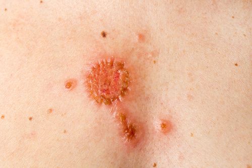
The first sign of basal cell carcinoma is usually a hardening of the skin. This can develop into a glassy-white to reddish nodule in which fine blood vessels are visible. In more advanced stages, weeping ulcers or crusts form on the affected area.
Symptoms of spinal cell carcinoma
Spinalioma also occurs mainly in areas that are often exposed to the sun, such as the bridge of the nose, forehead, ears, lower lip, neck, forearms, back of the feet and hands. The slowly growing cancerous tumor appears as a skin nodule or lump. It keratinizes over time and forms a crust. This can break open and then ooze or bleed.
Skin cancer – diagnosis at the USZ
In the event of a suspicious skin change, we will first obtain more detailed information about your medical history (anamnesis) during the initial consultation. For example, we ask when you first noticed the skin change, whether it has changed in any way since then (size, color) and whether there are any known cases of skin cancer in your family. Please also inform us of any pre-existing conditions and medication you are taking. This is followed by the following examinations, depending on the doctor’s assessment:
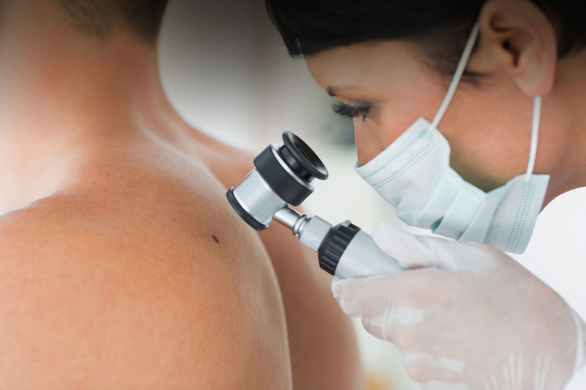
- Skin examination: We look at the skin all over the body, including all pigmentation spots and other abnormalities. We examine the suspicious skin change particularly carefully. A special microscope (dermatoscope or reflected light microscope) helps us to do this.
- Tissue sample: For a more detailed examination, we can take a tissue sample from this area (under local anesthesia). This is examined in the laboratory to determine whether it is actually skin cancer (or a precursor of it). In the case of minor skin changes, we may also cut them out completely and send them to the laboratory for analysis.
- Imaging procedures: If the laboratory confirms the suspicion of skin cancer, further examinations follow. This primarily includes imaging procedures such as X-rays, ultrasound, computer tomography and magnetic resonance imaging. They help to determine the exact location, extent and severity of the cancer. This is important for therapy planning.
Skin Tumor Center
At the USZ, numerous specialist departments have joined forces to form a skin tumor center. The center is certified according to the guidelines of the German Cancer Society (DKG). A team of experts specializing in the medical treatment of skin cancer works closely together here for the benefit of our patients. At DKG-certified centers, patients are treated according to strict quality criteria and, according to current studies, have a better chance of survival on average.
Skin cancer: prevention, early detection, prognosis
The best way to prevent skin cancer is to protect your skin from too much UV radiation. The most important recommendations are as follows:
- Stay in the shade between 11 a.m. and 3 p.m., when the sun is at its most intense.
- Protect yourself from excessive sunlight with a hat, sunglasses and long clothing (long sleeves, long pants).
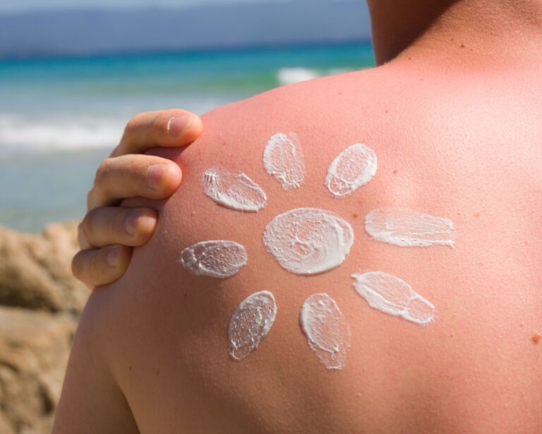
- Use sunscreen with a sufficiently high sun protection factor.
- Avoid visits to the solarium.
Take these recommendations to heart, especially if you have an increased risk of skin cancer – for example, because you have a fair skin type and/or there are already cases of skin cancer in your family.
Regular examinations by a dermatologist for the early detection of skin cancer can be useful for people with an increased risk of developing the disease. This is the case, for example, if someone has a large number (more than 100) of pigmentation marks on their body, has previously suffered from skin cancer or has an immunodeficiency (due to medication or illness). It is best to discuss with a doctor whether it would be advisable for you to have regular check-ups with a dermatologist.
Whether you have an increased risk of skin cancer or not – regular self-examination of your skin can’t hurt, so that you notice any suspicious skin changes at an early stage. Keep an eye on pigmentation marks in particular. If such a mark changes in size, shape or color, you should have this clarified.
Skin cancer – progression and prognosis
The earlier skin cancer is detected and treated, the better the chances of recovery.
- Black skin cancer: It is particularly important to treat black skin cancer in good time. It often grows slowly at first, but is nevertheless very dangerous. This is because once a melanoma has penetrated into deeper layers of the skin, individual cancer cells can easily spread throughout the body via blood and lymph channels and form metastases, for example in the lungs, liver, bones or brain. A cure then becomes difficult. If, on the other hand, malignant melanoma is detected early, when it is still confined to the epidermis, the chances of recovery are significantly better.
- Basal cell carcinoma: Basal cell carcinoma also grows very slowly, but almost never forms metastases. In advanced stages, however, it can expand in width and depth and subsequently destroy a lot of tissue (including bone and cartilage). Early treatment is therefore also advisable here. The chances of recovery are then very good. However, relapses (recurrences) occur more frequently after treatment has been completed – either in the same or a different part of the body.
- Spinal cell carcinoma: Spinal cell carcinoma also grows slowly and can usually be cured very well if treated early. However, if it is already more advanced, it can affect muscle and nerve tissue and form metastases (e.g. in lymph nodes, lungs, liver or bones). Then the prognosis worsens.
Second opinion for skin cancer
When a cancer diagnosis is made, a second medical opinion is an important decision-making tool. The Comprehensive Cancer Center Zurich supports you with a professional expert opinion. They receive a thorough analysis of the situation as well as personal advice and quick answers to their questions.
Skin cancer: treatment is planned individually
Various therapies are available for the treatment of skin cancer, including surgery, cold treatment, chemotherapy and radiotherapy. They can be combined in different ways, adapted to the individual patient. Decisive factors include the form and extent of the skin cancer, the location of the tumor and individual factors such as the age and general state of health of the person affected.
Interdisciplinary treatment
The USZ has a specialized team for each of the different types of skin cancer. A specialty of dermatology at the USZ is the removal of white skin cancer using so-called Mohs surgery: the tumor is cut out at a relatively close distance, after which the piece of tissue at the edges of the cut is immediately examined under a microscope. If tumor remnants are found, additional tissue is removed at this site. These steps are repeated until no more tumor remnants are found.
This procedure has two decisive advantages: Firstly, there are far fewer relapses: instead of around five to 25 percent of patients (depending on the type of tumor), only one to five percent have to be operated on again later due to a relapse. Secondly, the aesthetic result is better, as only as much tissue is removed as necessary.
If the white skin cancer is already so large that it can no longer be treated surgically or has already formed metastases (offshoots), medication or radiotherapy (or combinations) may also be necessary. Targeted therapy using smoothened inhibitors (tablets) is available for basal cell carcinoma, while immunotherapy with anti PD1 is now also approved for spinocellular carcinoma.


