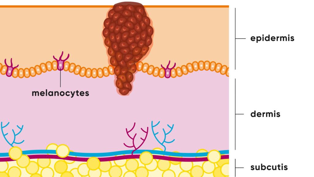
What is a melanoma
Melanoma, also known as malignant melanoma, is one of the most dangerous forms of skin cancer. It develops from the pigment-producing cells of the skin, the melanocytes. These produce the pigment melanin and are therefore responsible for tanning the skin. In melanoma, the melanocytes degenerate and multiply uncontrollably. In addition to melanoma of the skin, in rare cases melanoma can also develop on the mucous membranes inside the body or in the eye.
Melanoma types
Malignant melanoma is known for its many different manifestations, which can influence diagnosis and treatment. Early detection and correct classification of the different types of melanoma is crucial for effective treatment.
- Superficial spreading melanoma (SSM): The most common type of melanoma, known for spreading across the surface of the skin before growing deeper. It often shows a mixture of colors.
- Nodular melanoma: An aggressive type that develops quickly and is characterized by dark, raised nodules on the skin. This form can penetrate deep into the skin at an early stage and form metastases.
- Lentigo maligna melanoma: Occurs mainly in older people and develops from so-called lentigo maligna skin spots that have been exposed to the sun for a long time. They are typically flat and dark in color.
- Acrolentiginous melanoma (ALM): This type usually appears in areas that are difficult to observe, such as the palms of the hands, soles of the feet, and under the nails. It is rarer and can appear as a dark stripe or spot.
- Amelanotic melanoma: A particularly challenging form to identify, as it shows little to no pigmentation and often appears as a non-pigmented, raised lesion.
How does melanoma develop?
Melanoma is divided into different stages to describe the progression of the disease. These stages indicate how far the tumor has spread, whether lymph nodes are affected and whether metastases are present. The early stage of malignant melanoma marks the beginning of the disease, when the tumor is still small and has not spread widely.
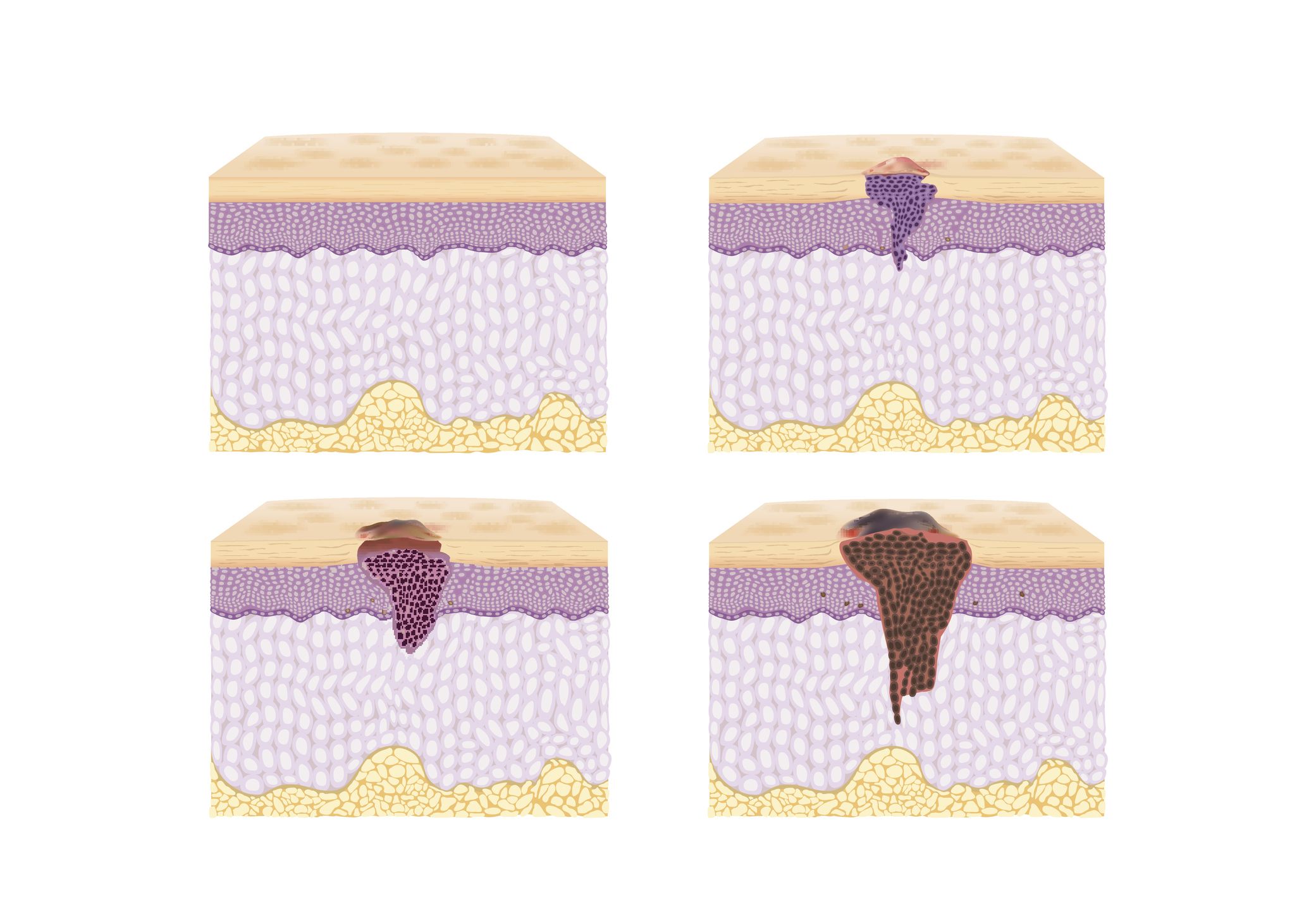
Simplified diagram showing the development of melanoma. The depth of penetration into the skin is decisive for the prognosis and further therapy.
- Top left: normal skin
- Top right: Preliminary stage of a melanoma in the uppermost layer of the skin (melanoma in situ)
- Bottom left: Melanoma that has broken through the basal layer of the skin
- Bottom right: deep melanoma extending into the subcutaneous fatty tissue
How common is melanoma?
Melanoma is the fourth most common type of cancer in Switzerland. In Switzerland, around 33 people per 100,000 are newly diagnosed with melanoma every year. In total, there are around 3,200 new cases per year in Switzerland. The number of new cases is increasing worldwide.
How dangerous is malignant melanoma?
In the majority of cases, melanoma can be cured with surgery. However, it can form metastases in other organs in around 15% of patients. This leads to life-threatening situations. The earlier a melanoma is discovered and removed, the better the chances of recovery. Cancer cells spread when they migrate via the lymphatic or blood vessels into the lymph nodes or internal organs and form new tumors there. The risk of this depends primarily on the thickness of the melanoma. The thicker (depth of penetration) the melanoma, the more likely it is to develop the ability to form offshoots. The faster the melanoma is treated, the thinner it is and the better the chances of survival.
Melanoma symptoms
In addition to the criteria set out in the ABCDE rule, the following symptoms also indicate a malignant melanoma:
- Itching, bleeding or weeping of the affected skin areas
- New development or significant growth of the pigmented mole
Furthermore, those affected can also suffer from various accompanying symptoms (so-called “B symptoms”), which can manifest themselves during the course of the disease. These include:
- Night sweats
- Unintentional weight loss
- Fever
However, these symptoms usually only occur in advanced stages of the disease.
What are the causes/risk factors for melanoma?
The main cause of melanoma is UV radiation. You are exposed to UV radiation when you are in the sun or in a solarium.
Particularly at risk are people who:
- have a total of more than 100 moles (liver spots) on the body
- have fair skin (skin type I-II, i.e. reddish-blonde hair, blue eyes or freckles and a quick tendency to sunburn)
- Have cases of melanoma in the family
- have a weakened immune system, e.g. after an organ transplant or with an immunocompromising disease such as HIV
- have already been diagnosed with melanoma in the past
- have suffered severe sunburns, especially in childhood and adolescence
How do you recognize a melanoma?
It is important that you check your skin regularly. For patients with light skin types and moles in particular, it is advisable to examine the skin yourself at three-monthly intervals for suspicious skin changes. If you are unsure, consult a doctor. Also look at the hairy scalp, the back of the ears, the back of the neck, the soles of the feet and the genital region, possibly with the help of a mirror.
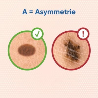
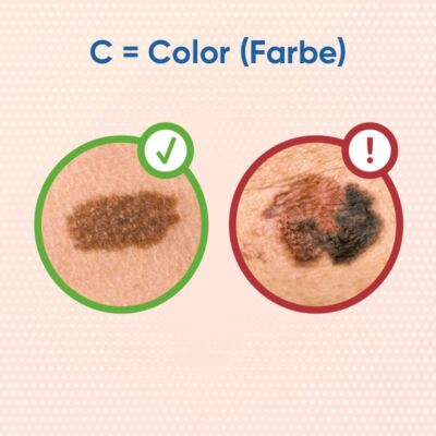
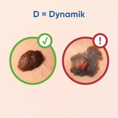
ABCD rule – skin cancer or birthmark?
The ABCD rule can help you to self-examine your skin in order to recognize changes at an early stage.
- A: Asymmetry: the dark skin spot is not circular, but uneven
- B: Irregular border: the skin patch is not sharply demarcated from the surrounding skin
- C: Color: the skin spot is rather dark brown to black
- D: Dynamics: the skin spot has changed in a relatively short time
Early detection
Moles that change in shape, color or size should be shown to a dermatologist immediately. This also applies to those moles that are different from the others, that itch or bleed from minor injuries. The “ugly mole principle” (moles that are obviously different from the other moles of the same person) can also help you to identify a conspicuous mole during self-examination, which should then be further clarified by a doctor.
Medical checkup – more information
Send a photo of the affected skin area and fill out the short questionnaire. Within 24 hours on weekdays, you will receive a reliable diagnosis from our experts. Your data will be transmitted to us encrypted and treated confidentially.
Early detection
The aim of early detection is to diagnose skin cancer, and melanoma in particular, at an early and therefore easily treatable or curable stage. At the University Hospital Zurich, we have a research focus on prevention and deal extensively with the early detection of skin cancer.
Diagnosis of melanoma
Suspicious moles or spots are usually removed completely under local anesthesia (excision). The tissue is then examined under a microscope (histology). Once the diagnosis of melanoma has been made, further examinations such as an ultrasound of the lymph nodes or a whole-body examination using PET-CT are carried out, depending on the depth of penetration of the melanoma.
Often the lymph node drainage is also examined and the sentinel lymph node is surgically removed. This also helps to identify small foci of offshoots, and further treatment is recommended if necessary. Your doctor will discuss the necessary examinations with you individually.
Melanoma treatment and therapy principles
The treatment depends primarily on the depth of penetration of the melanoma. So-called “thin” melanomas without offshoots can be cured with surgery in most cases. If the melanoma has already penetrated deeper into the skin or small foci of melanoma have been found in the lymph nodes, this is referred to as high-risk melanoma. Although they have not yet produced offshoots in distant organs, they do have a certain risk of doing so. If the melanoma has already spread to the organs, this is referred to as metastatic melanoma. The treatment approaches differ for these two forms.
In patients with high-risk melanomas, the tumor could be completely removed, but there is a certain risk of recurrent melanoma. The aim of treatment here is to achieve a permanent cure as a preventative measure. This is referred to as an adjuvant therapy approach.
In patients with organ infestation, a permanent cure can no longer be achieved in all cases. The aim is then to prevent the disease from spreading further and to maintain quality of life. However, even at this stage the disease can often be kept stable for years. This therapy principle is called palliative. The mainstays of treatment for melanoma are surgery, drug therapies and radiotherapy.
New opportunities through innovative therapies
Melanoma treatment has changed considerably in recent years. Immunotherapy has brought significant progress. Medication is used to stimulate the patient’s own immune cells so that they can recognize the cancer cells and fight against them. Around 40 percent of melanoma patients with immunotherapy respond to this treatment and benefit from its high efficacy. Even in advanced stages of the disease, a cure is possible with immunotherapy.
New therapeutic approaches for malignant melanoma
Support immunotherapy with stool transplantation
There is strong evidence from studies that the microbiome, i.e. the entire microorganisms in the gut, has an influence on whether patients respond well to immunotherapy. In order to harness this effect for patients with a weak response to the therapy, a research project at the USZ is transplanting prepared stool from patients who have responded well to immunotherapy to patients with a weak or no response. In a pilot study, it was shown that transplantation improved the effect of immunotherapies in these patients with advanced malignant melanoma.
Personalized therapy with molecular genetic analysis
Tumors are complex structures, the cancer cells interact with the cells in the surrounding tissue in various ways. Viewed at the cellular and molecular level, every tumor and every cancer is therefore unique – there is no such thing as “the” lung cancer or “the” melanoma. If these individual differences from patient to patient are taken into account during treatment, therapies can be used in a targeted manner and with the greatest effect. At the same time, for example, drugs that only have side effects and no medical benefit in the individual case are excluded and – conversely – the risk that a promising therapy is not used is reduced.
Such individually tailored treatment is possible thanks to molecular genetic analysis. For this purpose, a tissue sample of the tumor is analyzed in the laboratory using a special procedure. The information from this study makes it possible to determine which of the available cancer drugs is likely to be most effective in an individual case. This targeted and rapid use of the most suitable therapy opens up new opportunities for patients who were previously considered to be out of therapy.
More about cancer
Life expectancy and prognosis for malignant melanoma
Self-help group
An exchange with like-minded people can be very valuable. A self-help group for melanoma patients exists under the patronage of the Cancer League of the Canton of Zurich.
Life expectancy and prognosis for malignant melanoma
The life expectancy of malignant melanoma varies greatly depending on the stage of the disease at the time of diagnosis. Melanomas discovered at an early stage that are still confined to the upper layers of the skin have a very high 5-year survival rate of over 90%. This underlines the importance of early detection. In the case of more advanced malignant skin cancer, especially if metastases are already present, life expectancy decreases significantly.
However, the development of modern therapies such as targeted therapies and immunotherapies has improved treatment prospects and offers hope for a longer life even in advanced stages. Regular skin checks are therefore essential to increase the chances of a favorable prognosis
Black or white skin cancer?
Not all skin cancers are the same. Experts distinguish between white and black skin cancer, with the latter being referred to as malignant melanoma. White skin cancer can be divided into two main forms: Basal cell carcinoma and spinalioma, also known as squamous cell carcinoma.
Prevent skin cancer
Preventive measures are crucial to minimize the risk of skin cancer. Here are some important steps that everyone can take:
- Use sun protection: Regularly apply sun cream with a high sun protection factor, even on cloudy days.
- Seek shade: Avoid direct sunlight, especially during the peak hours from 10 am to 4 pm.
- Wear protective clothing: Long-sleeved shirts, pants and hats with wide brims offer additional protection.
- Use sunglasses: Protect your eyes and surrounding skin with glasses with UV protection.
- Examine skin regularly: Self-examination of the skin and regular checks by specialists.
- Be careful with sunbeds: Avoid the use of sunbeds and other sources of artificial UV radiation.
These measures can significantly reduce the risk of developing skin cancer.
Frequently asked questions about black skin cancer
Malignant melanoma can spread at different rates, often depending on the type and depth of the tumor. Metastasis can begin within a few months of the primary tumor developing.
The growth rate of melanoma can vary, some melanomas develop quickly within weeks to months, others grow more slowly.
Melanoma is particularly dangerous due to its ability to metastasize quickly and affect other organs, making treatment difficult and increasing the mortality rate.
Yes, melanoma, or malignant melanoma, is always malignant and poses a serious threat to health.