Research Group Sebastian Kozerke
Keywords
Medical Imaging, Cardiovascular Magnetic Resonance, Image Processing, Machine Learning, Digital Twinning
Summary & Mission statement
Development of diagnostic tools for quantification of blood flow, organ perfusion, metabolism and function, tissue composition, microstructure and mechanics.
The mission is to exploits principles from physics, electrical engineering and computer science to design highly efficient and sensitive imaging and spectroscopy approaches to help guide diagnosis and treatment.
Overview
We have developed space–time undersampling and parallel imaging techniques that significantly improve spatiotemporal resolution and scanning speed across a range of applications. These advances allow for more detailed and rapid evaluation of cardiac function, supporting earlier and more accurate diagnosis of cardiovascular conditions. Our research also encompasses diffusion MRI methods designed to investigate the microstructure of moving organs such as the heart. By visualizing microtissue structures at scales of just a few dozen micrometres, these techniques reveal key tissue properties essential for understanding and diagnosing cardiac disease.
In addition, we are advancing hyperpolarization technologies and related strategies to boost MRI signals by several orders of magnitude, enabling real-time metabolic imaging of the heart. Another major contribution is our development of variational network–based image reconstruction, an AI-driven approach that accelerates MRI processing by an order of magnitude. This improvement makes dynamic MRI of blood flow far more feasible in clinical settings. By delivering faster and more precise assessments of blood flow dynamics, this innovation holds strong potential for earlier detection and more effective treatment of cardiovascular disease.
Publications
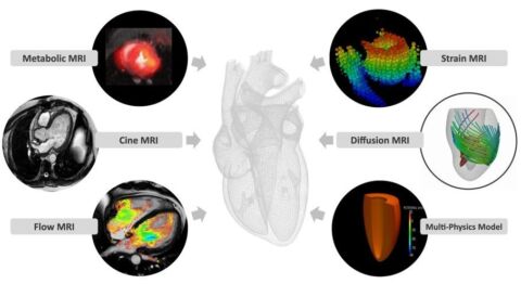
Based on MRI data acquired with newly developed methods, personalised digital twins can be created, i.e. patient-specific organ models that can be used to make disease predictions and virtual interventions possible.