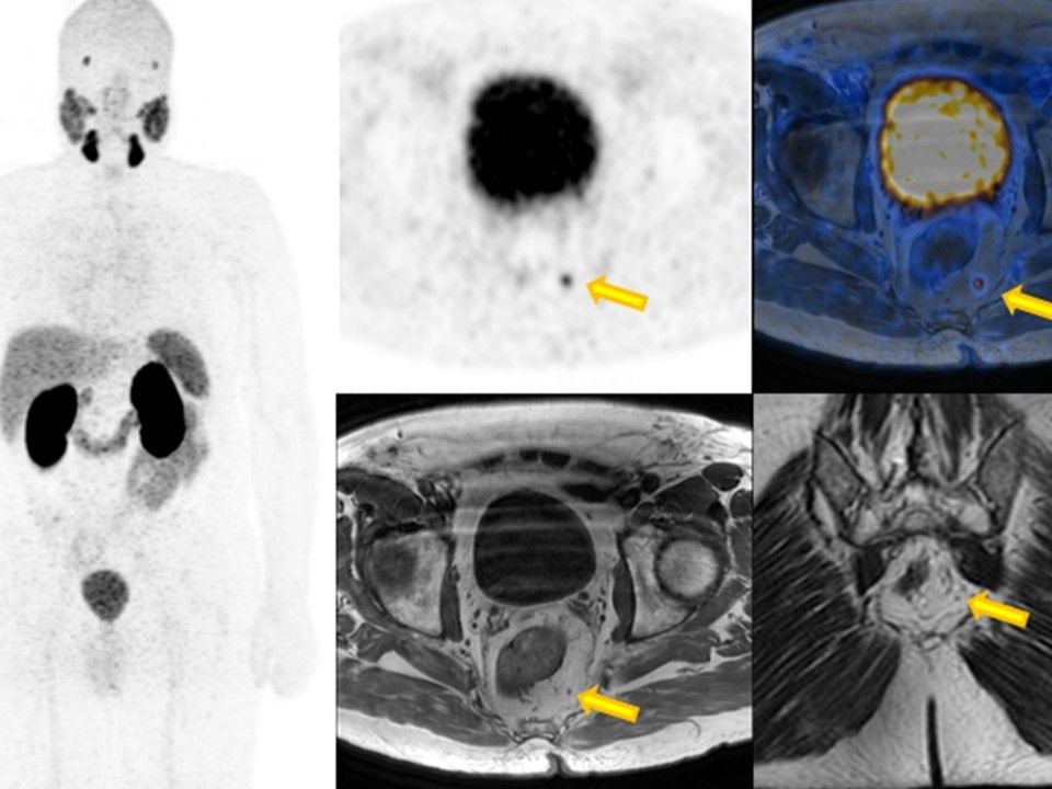
The coronary MIP image in this 67-year-old patient provides an initial overview. After a radical prostate resection in 2013, the PSA rose again to currently 0.22 ng/ml. On the axial fused images, a focal PSMA accumulation can be detected in a pararectal lymph node with a diameter of only 3 mm (yellow arrow). Despite its small size, this is therefore suspicious of a lymph node metastasis.