For patients
As a patient, you cannot register directly for a consultation. Please get a referral from your primary care physician, specialist.
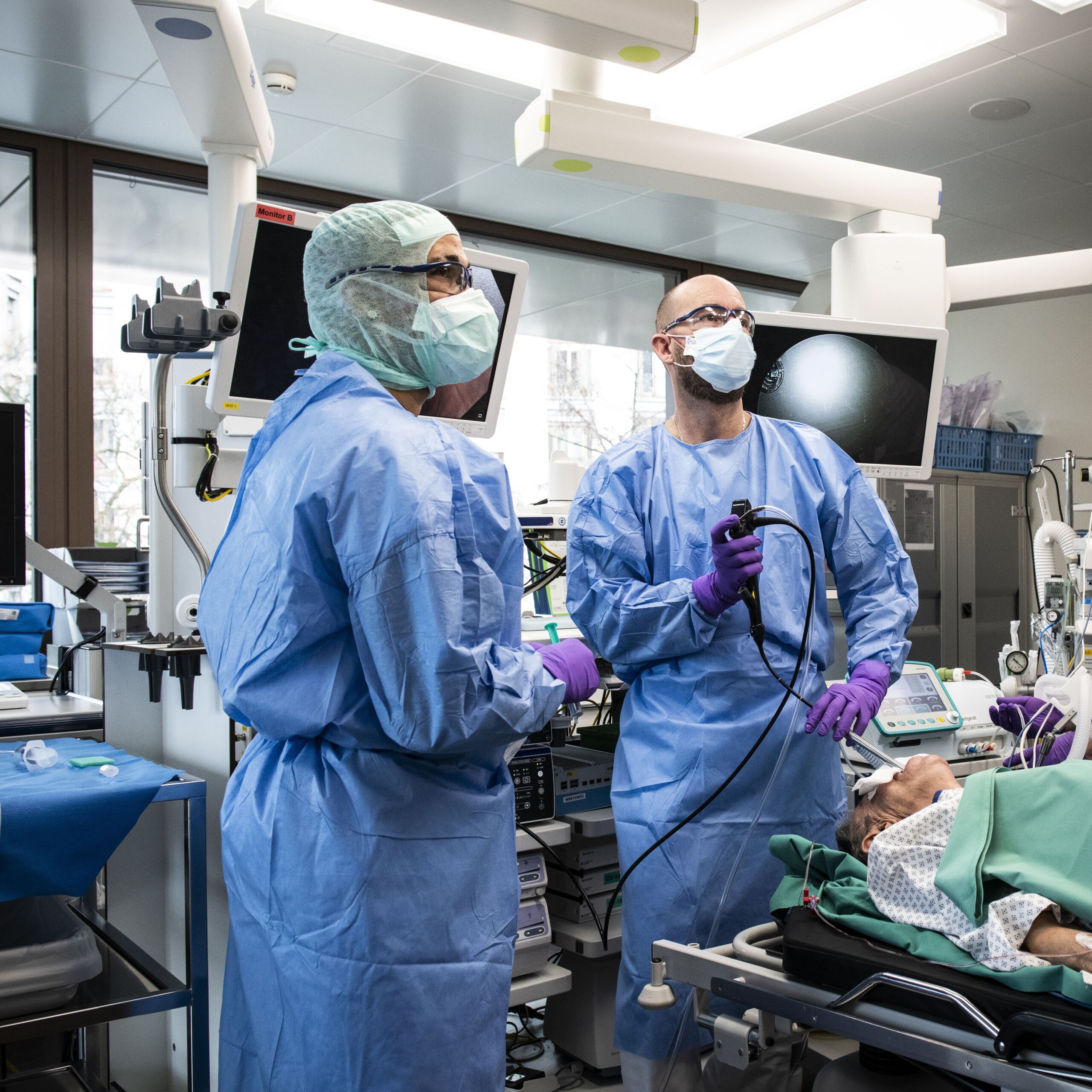
Interventional pneumology refers to all invasive procedures on the lungs and pleura. These include flexible and rigid bronchoscopy ("lung endoscopy") as well as pleural puncture and pleural drainage. In addition to diagnostic interventions, therapeutic procedures are also carried out on the lungs in order to favorably influence the course of the disease.
Bronchoscopy is used to diagnose and treat certain lung diseases.
Bronchoscopy for purely diagnostic purposes is primarily performed using a flexible instrument that is inserted into the airways via the oral cavity. Of course, this is done exclusively under sedation or general anesthesia, so that patients do not feel anything of the procedure. The main reasons for using a diagnostic bronchoscopy are suspected respiratory tract and lung infections, tumor diagnostics and the examination of enlarged lymph nodes in the respiratory tract. All modern methods of obtaining material for histological, cytological, microbiological and molecular biological examinations are available. In addition to X-ray fluoroscopy, ultra-thin bronchoscopes, endobronchial ultrasound (EBUS), electromagnetic navigation bronchoscopy and virtual bronchoscopy procedures are used for precise localization. In recent years, tissue preservation using a cryothermal probe has also become established for unclear lung diseases.
Patients must have an empty stomach for the bronchoscopy, regardless of whether the bronchoscopy is performed under sedation or general anesthesia. This means that no solid foods may be consumed within the six hours before the scheduled appointment. Drinking is permitted up to two hours before the procedure. Existing medication (especially blood-thinning medication) must be discussed with us. Depending on age, previous illnesses and the complexity of the procedure, bronchoscopy is performed on an outpatient or inpatient basis. Depending on this, the bronchoscopy is also performed either under sedation or general anesthesia.
The procedure itself only takes between 30 and 60 minutes.
The hospital stay is either outpatient or inpatient for two nights. Outpatients can work before the appointment without further ado (possibly half a day). After the bronchoscopy, the patient is unable to work on the day of the intervention. In addition, it is not permitted to drive a vehicle on the same day after the bronchoscopy. Patients are usually fully fit for work again the following day and their driving license is restored.
The follow-up treatment depends on the results of the examination. As a rule, patients are informed of the results of the examination by telephone or during a subsequent consultation, at which time treatment is also planned.
Bronchoscopy is a very low-impact, minimally invasive technology for the treatment of some respiratory and lung diseases.
Interventional bronchologyLung volume reduction is a potent and promising treatment method for patients with emphysema. Those affected suffer from severe shortness of breath at the slightest physical exertion, which can be attributed at least in part to over-inflation of the lungs. The aim of lung volume reduction is to reduce this overinflation. After the procedure, shortness of breath, quality of life, physical resilience and lung function improve. The University Hospital Zurich is one of the world’s leading centers for surgical and bronchoscopic lung volume reduction, with the Department of Pulmonology working closely with the Department of Thoracic Surgery. Several techniques are available for bronchoscopic lung volume reduction. First and foremost, small valves are inserted into the airways through the bronchoscope to “deflate” the lungs to a certain extent. Alternatively, bronchoscopic lung volume reduction can also be performed using steam or so-called coils.
Overview of current methods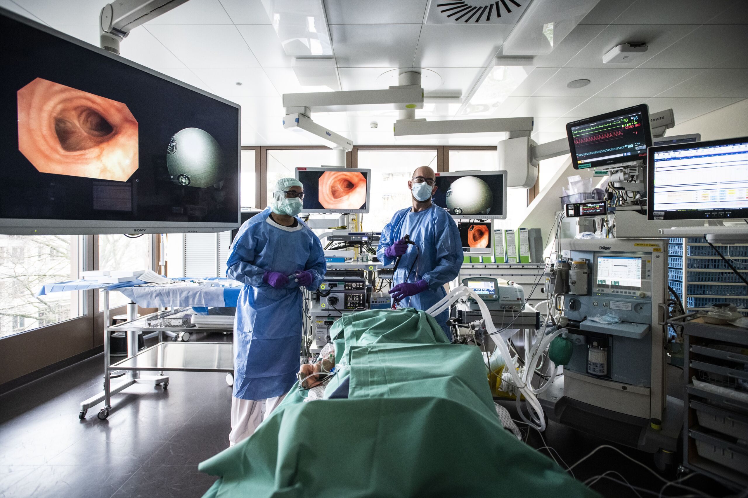
Patients undergoing bronchoscopic lung volume reduction are usually admitted to hospital one day before the procedure so that certain preliminary examinations, including visits by the anesthetist, can be carried out promptly. The procedure itself is always performed under general anesthesia and takes about 60 minutes.
The hospital stay is inpatient and patients usually stay there for five to seven nights. In principle, it is possible to return to work after the inpatient stay. However, experience shows that a gradual increase in workload makes more sense.
We recommend outpatient or inpatient pulmonary rehabilitation after lung volume reduction. This can further improve the positive effects.
If the airways are affected by malignant tissue (e.g. lung cancer), therapeutic bronchoscopy offers various options for clearing the airways and thus improving breathing. In most cases, modern and gentle techniques are used during a rigid bronchoscopy (argon plasma coagulation, diode laser, cryothermal probes, diathermy probes and dilatation balloons). It may also be necessary to insert a stent into the airways. Bronchoscopy is frequently used for diagnostic and therapeutic purposes in cases of pulmonary bleeding (hemoptysis or hemoptysis). In addition to laser, argon plasma or the administration of hemostatic medication directly via the bronchoscope, a bronchus blocker can be inserted to isolate the bleeding section of the lung, depending on the location and severity of the bleeding.
Another focus of therapeutic bronchoscopy is the treatment of benign (idiopathic) tracheal stenoses (dilatation tracheoplasty) and the treatment of pulmonary alveolar proteinosis using whole lung lavage.
For a rigid bronchoscopy, patients are usually admitted to hospital as inpatients one day before the procedure so that certain preliminary examinations, including visits by the anesthetist, can be carried out promptly. The procedure itself is always performed under general anesthesia and usually takes 60 to 90 minutes.
The hospital stay is inpatient and usually lasts three to four nights. In principle, it is possible to start work after the inpatient stay. However, our experience shows that a gradual increase in workload makes more sense.
The follow-up treatment depends on the underlying disease. In the case of cancer, further treatment is discussed and coordinated at the Interdisciplinary Tumor Board. In the case of benign tracheal stenoses, an outpatient follow-up consultation is usually scheduled four to six weeks after the procedure.
A further spectrum of interventional pneumology includes diagnostic and therapeutic procedures for pleural effusions and pneumothorax. In the case of malignant pleural effusions, a pleural catheter that remains in the skin (PleurX catheter) can also be inserted under local anesthesia. This can also be removed after a few weeks or months in some cases.
Senior Physician, Department of Pulmonology
As a patient, you cannot register directly for a consultation. Please get a referral from your primary care physician, specialist.
Assign your patient online, by e-mail or by post.
University Hospital Zurich
Department of Pulmonology
To the attention of (insert desired doctor here)
Raemistrasse 100
8091 Zurich