Alongside self-examination, mammography is the most important method for the early detection of breast cancer. The University Hospital Zurich uses the only spiral computer tomograph in Switzerland specially developed for breast examinations. A study has now confirmed the high level of acceptance among patients as well as the high image quality and user-friendliness.
In addition to self-examination, mammography is the most important method for the early detection of breast cancer. Many patients find conventional X-ray examinations using mammography or tomosynthesis unpleasant or even painful. Some women therefore delay the upcoming appointment or don’t go for a check-up at all.
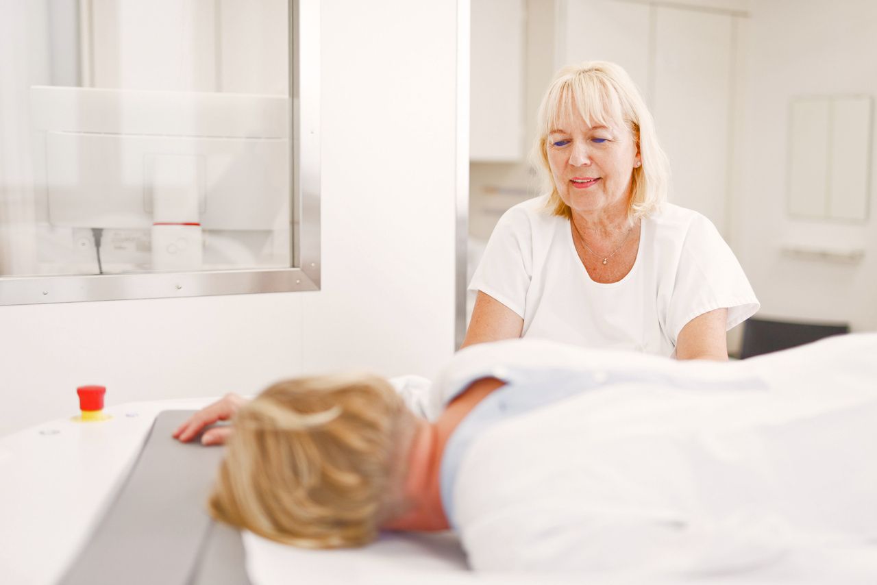
Since 2018, the Institute of Diagnostic and Interventional Radiology at the USZ has also been offering mammograms with a spiral computed tomography device – the only one of its kind in Switzerland. With breast CT, mammograms can be taken without putting pressure on the breast, as the examination is carried out lying down and not standing up. A state-of-the-art photon-counting detector is also used. In a retrospective study, the comfort for patients during the examination has now been assessed and the user-friendliness and image quality evaluated. For this purpose, 2,418 B-CT scans of 1,222 women were analyzed. The patients rated their comfort during the examination, the radiology assistants rated the patient’s freedom of movement and the operability of the device, while the radiologists rated the lesion contrast, the detectability of calcifications, the complete imaging of the breast and the overall image quality.
The breast CT impresses patients and users alike
The evaluation of the data confirmed the everyday experience in practice:
- The patients rated the comfort very highly. More than 99% reported “no” or “negligible” complaints during the examination.
- The patients’ freedom of movement and the user-friendliness of the breast CT were each rated as “no” or “negligible” discomfort in >99%.
- The picture quality was rated as “none” or “negligible limitations” in 96.7% of cases.
- The lesion contrast and detectability of calcifications were rated as “optimal” or “good” in 92.6% and 98.4% respectively.
- A “complete” and “almost complete” image of the breast was found in 41.9%, while the pectoral muscle was not covered in 56.0% of the images. In 2.1% of cases, larger parts of the breast were not imaged.
Normally, the detection rate for a mammogram is 0.4%. If an ultrasound were always performed in addition for dense breast tissue, the detection rate would improve to approx. 0.7%. If an additional ultrasound examination is always performed in patients with dense breast tissue, the detection rate for breast CT is 1.4% according to internal evaluations.
Professor Thomas Frauenfelder, Head of the Institute of Diagnostic and Interventional Radiology at the USZ, explains the significantly better result primarily by the fact that, thanks to the painless examination, women who have not had a mammogram for years or have never had one can also be examined using breast CT. “Especially for women who have had bad experiences with conventional mammography or are afraid of it, breast CT is the alternative.”
Mammographie Screening
The Institute of Diagnostic and Interventional Radiology has numerous imaging techniques for visualizing breast tissue. The most suitable method is used depending on the clinical problem.
No additional costs for a breast CT
The costs covered by health insurance for a breast CT are the same as for a conventional mammogram with tomosynthesis. The preventive examinations, which are not yet covered in some cantons (including the canton of Zurich) in contrast to the diagnostic examinations, are therefore not associated with additional costs. In addition to breast CT examinations, the USZ continues to offer the classic procedure.
Link to publication: Clinical assessment of image quality, usability and patient comfort in dedicated spiral breast computed tomography. DOI: 10.1016/j.clinimag.2022.07.001