
The purpose of this work was to further analyze concentration-dependent effects of Dutasteride on PSMA expression in a mouse xenograft model.
Cancer Rep (Hoboken). 2021 Dec;4(6):e1418.
Kranzbühler B, Sousa R, Prause L, Burger IA, Rupp NJ, Sulser T, Salemi S, Eberli D
*CCCZ Members in bold
Abstract
Background:
Dutasteride has been shown to increase expression of the prostate-specific membrane antigen (PSMA) in prostate cancer cells in previous in vitro studies. This 5-alpha-reductase inhibitor is commonly used for the treatment of symptomatic benign prostatic enlargement. The modulation of PSMA expression might affect PSMA-based prostate cancer imaging and therapy.
Aim:
The purpose of this work was to further analyze concentration-dependent effects of Dutasteride on PSMA expression in a mouse xenograft model.
Methods and results:
Four groups of mice bearing LNCaP xenografts were treated for 14 days with daily intraperitoneal injections of either vehicle control or different concentrations of Dutasteride (0.1, 1, 10 mg/kg). Total expression of PSMA, androgen receptor (AR), and caspase-3 protein was analyzed using immunoblotting (WES). In addition, PSMA, cleaved caspase-3 and Ki-67 expression was assessed and quantified by immunohistochemistry. Tumor size was measured by caliper on day 7 and 14, tumor weight was assessed following tissue harvesting. The mean PSMA protein expression in mice increased significantly after treatment with 1 mg/kg (10-fold) or 10 mg/kg (sixfold) of Dutasteride compared to vehicle control. The mean fluorescence intensity significantly increased by daily injections of 0.1 mg/kg Dutasteride (1.6-fold) as well as 1 and 10 mg/kg Dutasteride (twofold). While the reduction in tumor volume following treatment with high concentrations of 10 mg/kg Dutasteride was nonsignificant, no changes in AR, caspase-3, cleaved caspase-3, and Ki-67 expression were observed.
Conclusion:
Short-term Dutasteride treatments with concentrations of 1 and 10 mg/kg significantly increase the total PSMA protein expression in a mouse LNCaP xenograft model. PSMA fluorescence intensity increases significantly even using lower daily concentrations of 0.1 mg/kg Dutasteride. Further investigations are needed to elucidate the impact of Dutasteride treatment on PSMA expression in patients.
Keywords:
Dutasteride; prostate cancer; prostate-specific membrane antigen
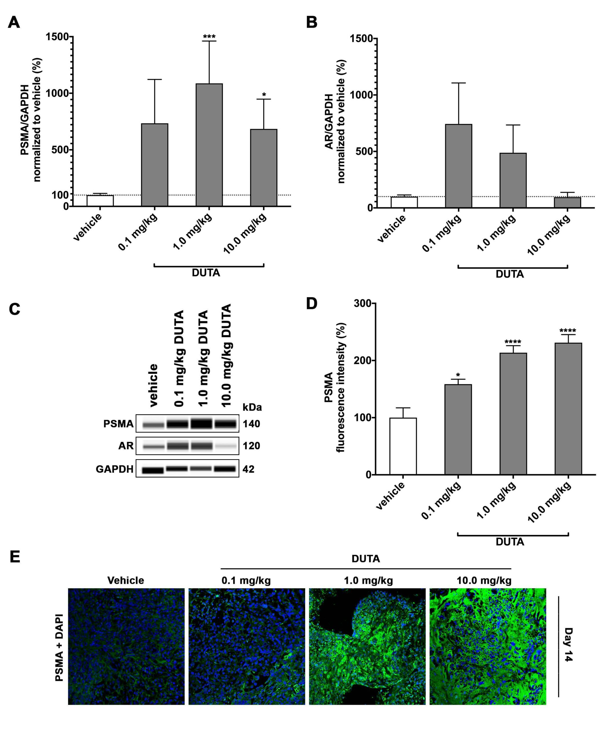
Figure 3 – PSMA and AR expression: Total PSMA protein expression (A) and total AR protein expression (B) was measured on harvested tumor tissue from mice treated with daily intraperitoneal injections of vehicle, 0.1 mg/kg Dutasteride, 1.0 mg/kg Dutasteride and 10.0 mg/kg Dutasteride for 14 days. PSMA and AR expression is presented as percentage compared to vehicle control. Data is shown as mean ± standard error of the mean (SEM) of three to six independent experiments. *p<0.05, **p<0.01, ***p<0.001, ****p<0.0001. Protein simple immunoblotting (WES) from one representative experiment (C). PSMA fluorescence intensity was measured and quantified using image J software. The results are presented as percentage of PSMA expression compared to vehicle control (D). Visualization of PSMA expression using immunohistochemistry (E). Representative confocal images of PSMA surface staining. Tumor tissue sections were stained with primary anti-PSMA antibody and detected using FITC (green) conjugated secondary antibody and DAPI (blue, 4′,6-diamidino-2-Phenylindole).