VECTRA® 3D whole-body photography is a modern technology for advanced skin cancer screening at the Dermatology Clinic of the University Hospital Zurich. The procedure enables a standardized, three-dimensional and high-resolution recording of the entire skin surface within a few seconds.
The method is particularly helpful for patients with an increased risk of black skin cancer (melanoma), for example if they have many or atypical nevi (moles), a personal or family history of melanoma or a genetic predisposition. The combination of whole-body imaging and sequential digital dermoscopy (close-up images) allows skin changes to be precisely documented and assessed over time.
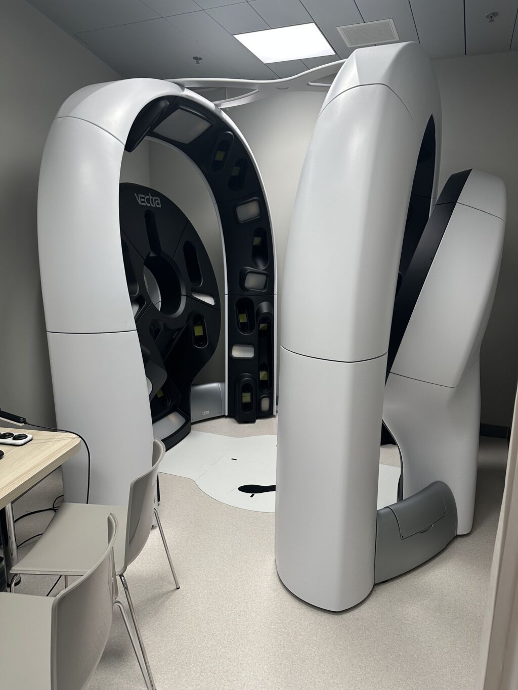
What is VECTRA?
VECTRA® is a 3D whole-body photography system that captures the skin surface in a standardized, three-dimensional and high-resolution way. The imaging serves as digital initial documentation for subsequent follow-up checks.
The method does not replace a medical examination, but it does complement it. Abnormal lesions or lesions that need to be checked can also be documented dermatoscopically and specifically compared over time.
What does digital progress monitoring do?
Longitudinal assessment using whole-body images makes it possible to detect new or morphologically changing lesions at an early stage. As a result, malignant transformations – the development of malignant melanoma – can be detected more quickly and unnecessary excisions can be avoided.
Published data from our group show that 3D whole-body imaging can achieve high diagnostic performance in the early detection of melanoma under real-world conditions [1]. In a Swiss survey, the majority of high-risk patients preferred the combination of 3D whole-body photography and medical supervision over clinical examination alone, or supplementary 2D whole-body photography or isolated AI approaches [2].
What does AI-supported risk assessment mean?
In addition to the medical examination, an AI-supported assessment can be used for selected moles. To do this, individual skin changes are photographed in greatly magnified form using digital dermoscopy. The AI analyzes these detailed images and provides a risk assessment that complements the medical assessment. However, the final decision is always made by the attending physician.
Where are the limits?
3D full-body photography and the optional AI-supported risk assessment are very helpful additional tools, but are no substitute for clinical expertise or dermoscopy. Conspicuous, new or changing lesions must continue to be assessed by a doctor.
In case of suspicion, histologic examination after excision or biopsy remains the diagnostic reference standard.
Frequently asked questions about VECTRA
No. The examination is non-invasive and does not require the removal of tissue. The image capture itself only takes a few seconds.
No. VECTRA complements the clinical examination and dermoscopy. Particularly relevant or conspicuous lesions are also documented dermatoscopically and specifically assessed in the course of the procedure.
The method is particularly suitable for patients with an increased risk of melanoma, for example with previous melanoma, a family history of melanoma, a genetic predisposition, many nevi or atypical or dysplastic nevi.
In suitable situations, structured follow-up can help to better distinguish stable lesions from new or changed lesions. The aim is to reduce unnecessary excisions and detect relevant changes at an early stage.
The AI assesses enlarged detailed images of individual moles and provides an additional assessment of the risk. The result is a decision-making aid for the doctor, not an automatic finding and not a substitute for a personal examination.
The costs for up to two full-body photography examinations per year are generally covered by health insurance.