Pseudarthrosis (failure of bone fractures to heal) Therapy
Modern concepts for the treatment of pseudarthrosis
Bone healing is multifactorial and dependent on:
Maintained blood circulation
Vital bone cells: New bone formation
Sufficient stability after bone fractures
Definition of pseudarthrosis and example of pseudarthrosis
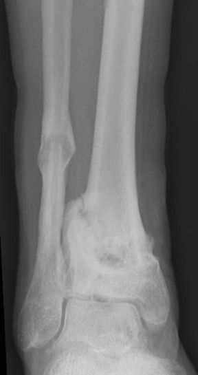
X-ray of an open fracture
The x-rays show a gap formation as an expression of an unhealed bone fracture = pseudarthrosis (false joint formation) just above the upper ankle joint.
Case study
In principle, there is a high risk of pseudarthrosis developing after an open fracture.
X-ray of an open fracture
This is due to an injury-related circulatory disorder:
Open fracture
Severe soft tissue injury
There are other risk factors for the development of pseudarthrosis
Risk factor instability of the osteosynthesis for the development of pseudarthrosis
Case studies
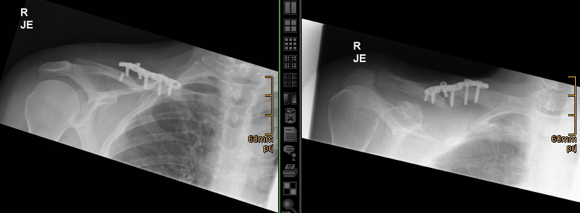
Avulsion and fracture of a plate osteosynthesis on the collarbone
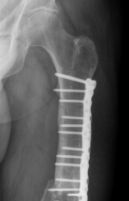
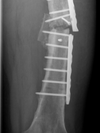
Broken plate in the thigh
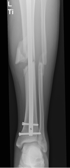
Broken plate in the lower leg
A typical symptom of pseudarthrosis is often present
Exercise-induced pain
Swelling
False joint mobility
X-ray diagnostics
Case study
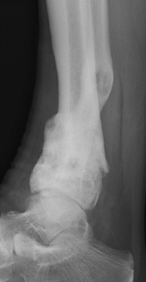
Conventional X-ray diagnostics for the detection of pseudarthrosis is not always clear!
X-ray images of the lower leg. Has the bone healed?
X-ray images of the lower leg. Has the bone healed?
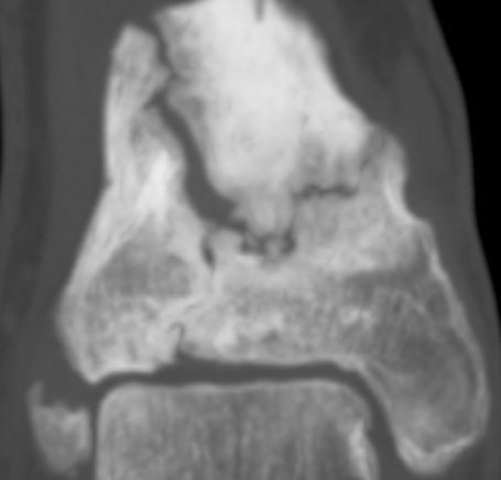
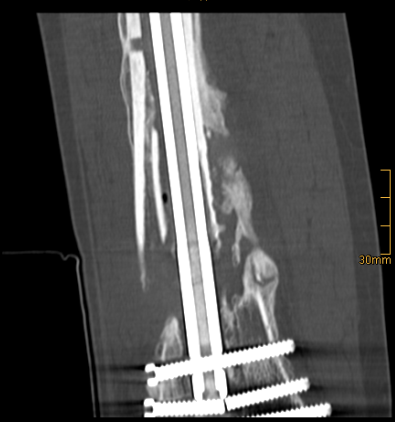
Extended diagnostics with tomographic imaging of the pseudarthrosis region using computer tomography shows findings typical of pseudarthrosis
The CT images show a bone gap and a screw fracture
The following therapy principles are used to treat pseudarthrosis
Increased stability:
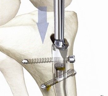
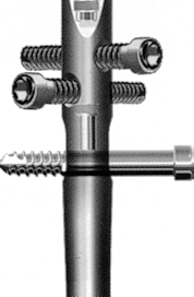
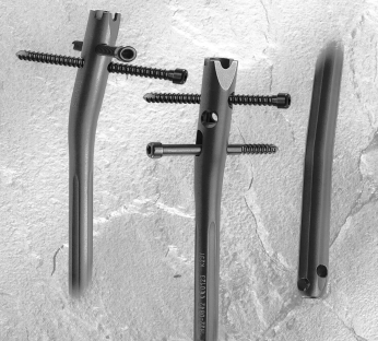
nail filling the medullary canal
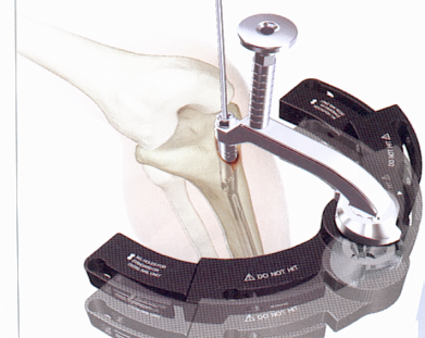
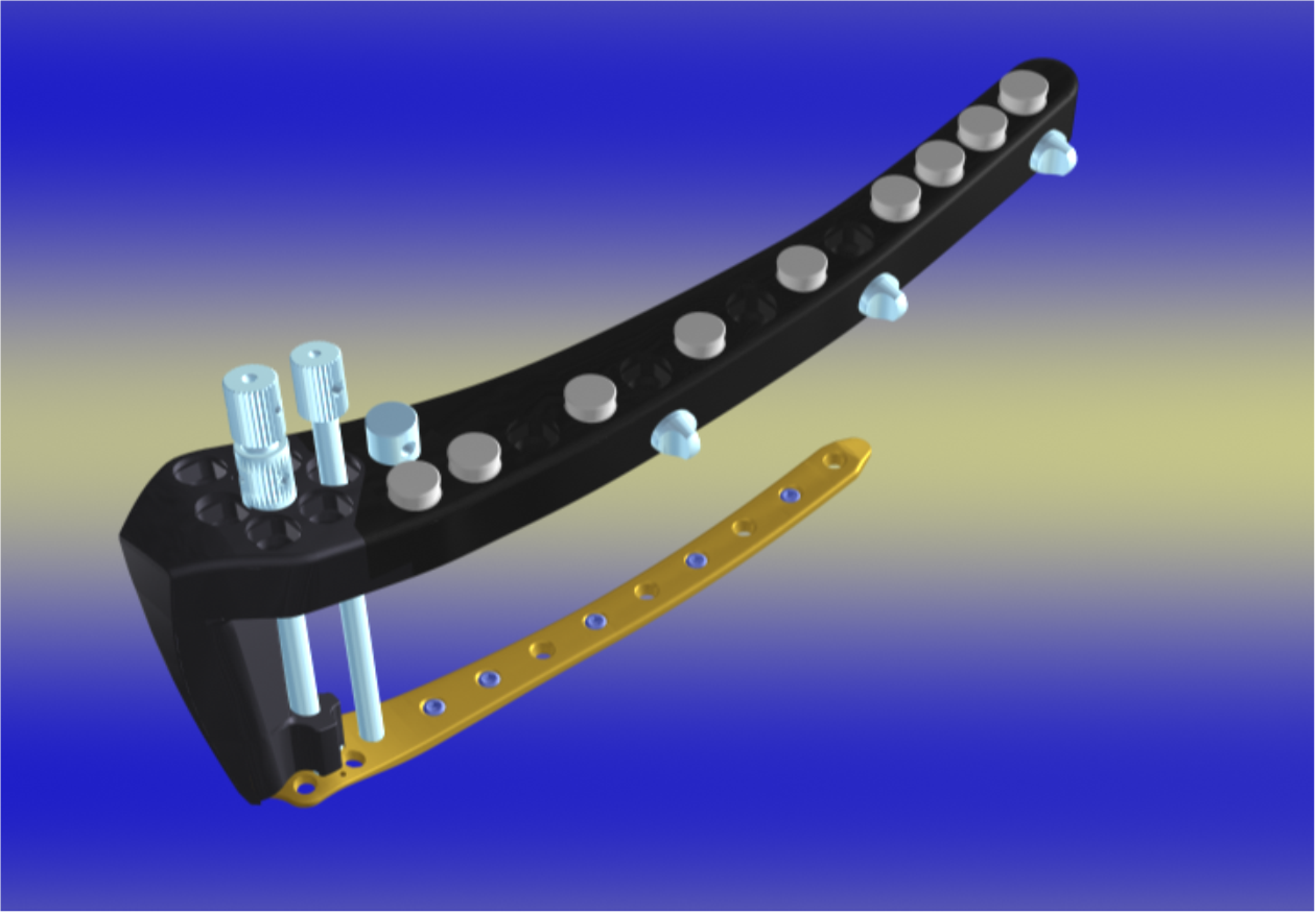
Angle-stable, bridging plate
Correction of bone misalignment
Biological activation
Implants to stabilise the bone: nail filling the medullary cavity
Implants to stabilize the bone: Modern angular stable, bridging plate
Case studies for the surgical treatment of pseudarthrosis
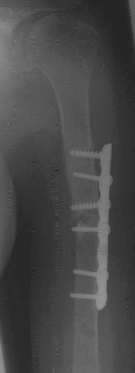
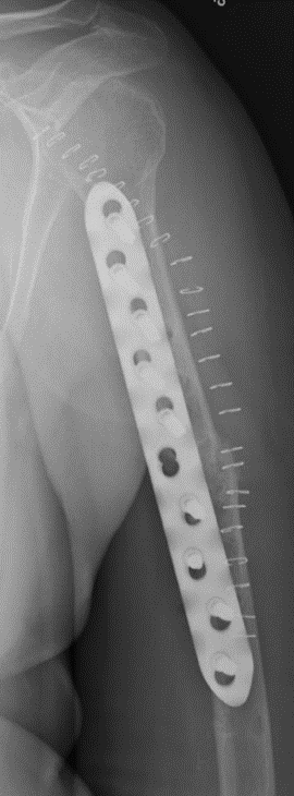
The X-ray analysis of a humeral shaft pseudarthrosis after plate osteosynthesis shows the following findings:
Plate and screws are loose
The broken bone has not healed
The bone is unstable
Operation:
Removal of the loosened plate
Resection of scarred connective tissue that prevents new bone formation
Change to stable-angle, long-span, bridging plate osteosynthesis
Accumulation of autologous bone and bone growth factor BMP-2
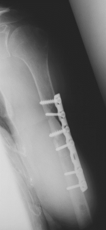
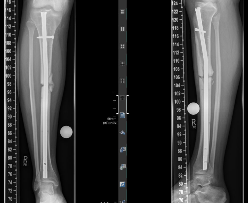
The X-ray analysis of a tibial pseudarthrosis after intramedullary nailing shows the following findings
The broken bone has not healed
The inserted intramedullary nail is unstable and the locking screws remote from the body have been removed
A standardized surgical treatment for lower leg pseudarthrosis consists of changing the intramedullary nail:
Drilling out and thus “freshening up” the medullary cavity
Implantation of a stable intramedullary nail
Compression of the pseudarthrosis zone
Locking screws inserted away from the body increase stability
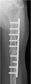
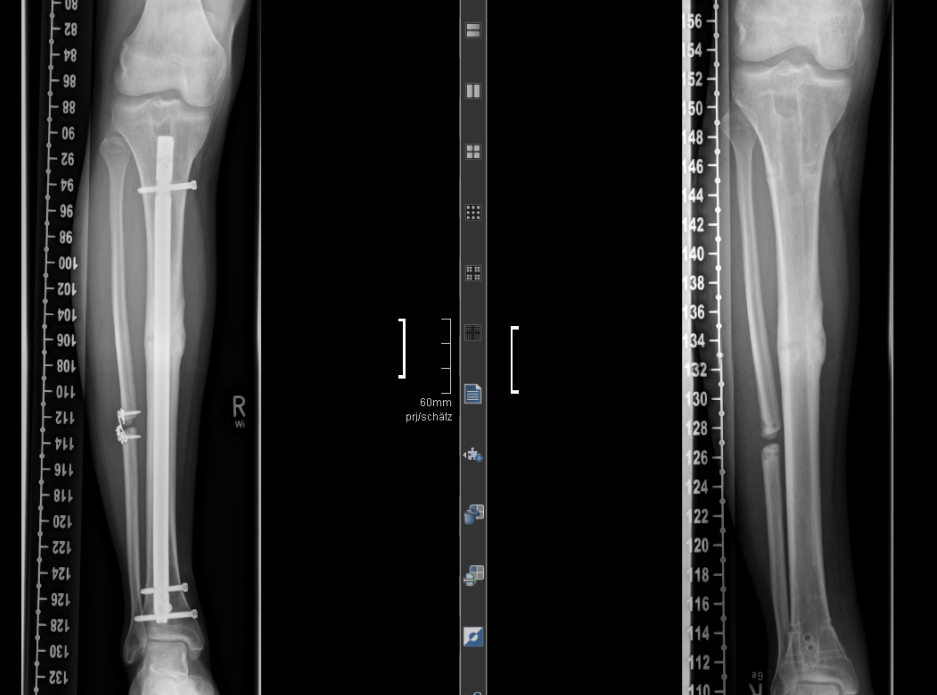
The picture already shows the complete bony healing of the former pseudarthrosis after 3 months
The picture on the right shows the metal removal after approx. 1 year
The following procedures are used for the biological activation of the pseudarthrosis region
Transplantation of bone from the iliac crest
Accumulation of bone growth factors
Blood collection from the iliac crest
Another option for harvesting autologous bone is the so-called RIA system
Removal of bone cancellous bone from the bone canal, e.g. on the thigh
Summary: Modern treatment of pseudarthrosis (failure of bone fractures to heal)
Early surgical treatment of pseudarthrosis is recommended
Axle correction
Stabilization with
Most modern intramedullary nails
angle-stable plates
Biological activation of the pseudarthrosis zone with
Autologous bone
Bone growth factors support the successful healing of pseudarthrosis
A supportive therapy for pseudarthrosis
e.g. with high-energy shock waves can be considered in individual cases
For patients
As a patient, you cannot register directly for a consultation. Please get a referral from your primary care physician, specialist. For questions please use our contact form.