Accurately assessing an individual’s risk of developing the disease and determining the best possible treatment requires modern imaging technology and the expertise of the treating physicians. Therefore, such vascular malformations should be treated at centers that have the necessary experience and infrastructure.
Causes
The causes depend on the specific vascular malformation. Many arteriovenous malformations and cavernomas are congenital, although thereare also acquired forms1. In contrast, dural arteriovenous fistulas often do not develop until later in life. Possible causes of dAVF include head injuries, previous surgeries, inflammation, or blockages in cerebral veins. However, it is not always possible to identify a clear cause.
Symptoms of Vascular Malformations
Many vascular malformations do not cause any symptoms for a long time. If symptoms occur, they can vary greatly depending on the location and nature of the change.
Possible symptoms include:
- Headaches or dizziness
- Epileptic Seizures
- Weakness or paralysis
- Sensory disturbances
- Speech or vision problems
- Balance and Coordination Problems
- Concentration and Memory Problems
- Pulsating tinnitus (in certain cases of dAVF)
One risk associated with these vascular malformations is the occurrence of bleeding in the brain or spinal cord. This condition often manifests as a sudden, severe headache, neurological deficits, or impaired consciousness, and constitutes a medical emergency.
What types of vascular malformations are there?
Different vascular malformations can present in very different ways. The key factor is how the blood vessels are affected and where they are located. This determines the possible symptoms, the risk, and the appropriate treatment.
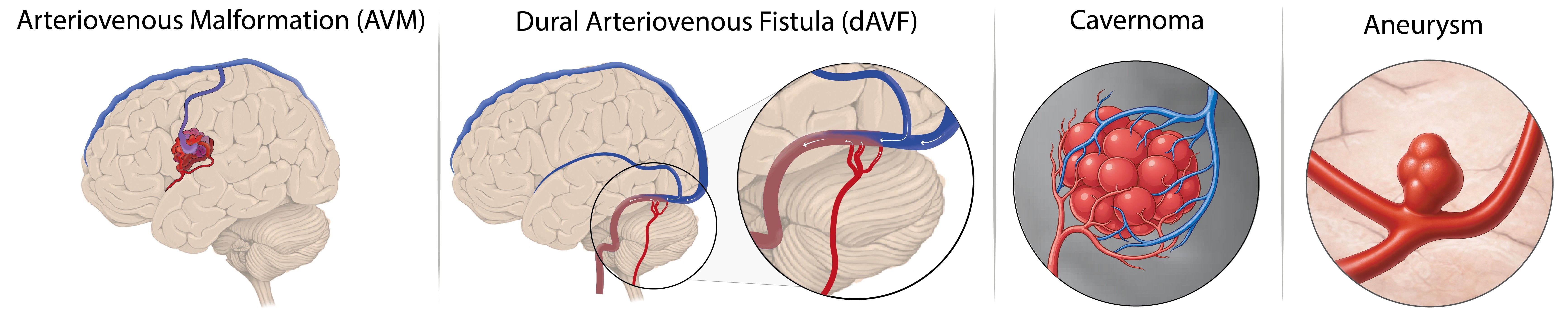
Figure 1: Vascular malformations of the brain and spinal cord. This figure schematically illustrates the four most common vascular malformations of the brain and spinal cord. From left to right: arteriovenous malformation (AVM), dural arteriovenous fistula (dAVF), cavernoma, and cerebral aneurysm.
Generally speaking, we distinguish between the following major types of vascular malformations:
- Arteriovenous Malformation (AVM): An AVM is a malformation characterized by an abnormal direct connection between inflowing and outflowing blood vessels. This results in a tangle of blood vessels through which blood flows at a higher pressure.
- Dural arteriovenous fistula (dAVF): In a dAVF, there is also a direct connection between the afferent and efferent blood vessels, through which blood flows at elevated pressure. Unlike an AVM, this malformation does not form a tangle of blood vessels; furthermore, a dAVF is not located in the brain or spinal cord itself, but rather in the protective meninges that surround the nervous system.
- Cavernoma – A cavernoma consists of small, dilated blood vessels through which blood flows slowly. It may remain asymptomatic for a long time, but it can also cause symptoms or bleeding.
- Brain aneurysm – A brain aneurysm is a balloon-like bulge in the wall of a blood vessel in the brain. Many brain aneurysms go unnoticed for a long time and do not cause any symptoms. However, depending on its size, shape, and location, there may be an increased risk that the aneurysm will rupture and cause a life-threatening brain hemorrhage.
More Information About Brain Aneurysms
Below, we explain each of these vascular malformations individually, describe typical symptoms, and outline the most important diagnostic tests.
Arteriovenous Malformation (AVM)
An arteriovenous malformation, or AVM for short, is a rare and specific malformation of the blood vessels in the brain or spinal cord. Normally, blood flows from the incoming vessels (“arteries”) through a very fine network of tiny vessels (“capillaries”) and slowly continues on to the outgoing vessels (“veins”). In this process, the high pressure from the incoming blood vessels is gradually reduced as the blood passes through the capillaries and is then transported at low pressure through the veins. In an AVM, this protective network of capillaries is missing. Instead, there is a direct connection between the afferent and efferent vessels, which form a cluster of vessels (“nidus”). Because of this direct connection, the blood flows significantly faster and at a higher pressure than intended. The draining vessels are often not designed for this and can become overloaded over time.
This can result in reduced blood flow to the surrounding brain tissue or cause blood vessels within the AVM to rupture, which can lead to a life-threatening brain hemorrhage. The average risk of bleeding from an untreated AVM is about 2–4% per year, but it canvary significantly depending on the AVM’s structure and location.²
A brain hemorrhage is a potentially life-threatening condition that often occurs suddenly and can cause neurological deficits. After a previous episode of bleeding, the risk of further bleeding is significantly increased, especially during the firstyear.³
The symptoms that occur and the risk of bleeding depend primarily on the size, shape, and location of the AVM.
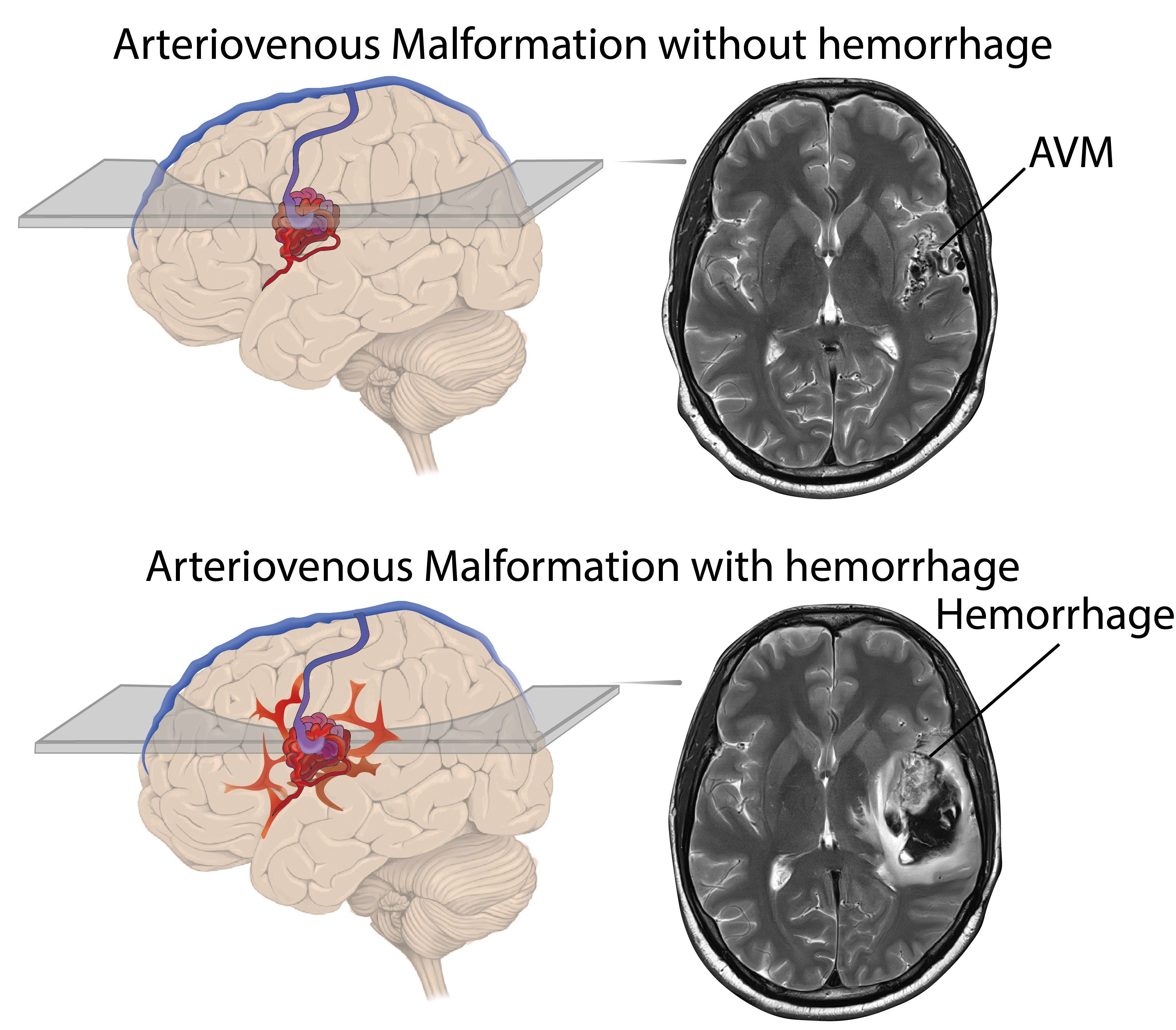
Figure 2: Arteriovenous malformation. On the left is a schematic representation of a brain with an AVM; on the right is the corresponding MRI cross-sectional image. The image above shows an AVM with no signs of bleeding. The image below shows the same AVM with typical signs of a dangerous brain hemorrhage. The figures are modifiedfrom 1.
What symptoms can an AVM cause?
The symptoms that may occur depend on the location of the AVM and the extent to which blood flow is altered. Some AVMs do not cause any symptoms for a long time and go unnoticed. In other cases, various symptoms may occur.
Common possible symptoms include:
- Neurological deficits, for example
- Weakness or signs of paralysis
- Sensory disturbances
- Speech or vision problems
- Balance or coordination problems
- Epileptic Seizures
- Problems with concentration or memory
- Headaches, dizziness, and other symptoms
In some cases, bleeding in the brain may occur. This condition typically manifests as sudden, severe headaches, neurological deficits, or altered consciousness, and constitutes a medical emergency.
Not every AVM causes symptoms, and not every AVM requires immediate treatment. It is therefore essential that medical experts conduct a thorough evaluation to accurately assess an individual’s risk.
Imaging tests are necessary to reliably detect an AVM and accurately assess the risk. These images show where the AVM is located, how large it is, and how blood flows through the vessels.
How is an AVM diagnosed?
The following tests are used for diagnosis:
- Magnetic Resonance Imaging (MRI): This test provides a very detailed image of the brain and spinal cord, as well as their blood vessels. An MRI helps to visualize the location and extent of the AVM, as well as its potential impact on the surrounding tissue.
- Computed Tomography (CT): A CT scan can quickly and reliably detect bleeding in the brain and spinal cord, which is why it is often used in emergency situations.
- Catheter angiography (DSA): In this procedure, a very thin tube is guided through a blood vessel in the arm or leg to the blood vessels in the brain. Contrast agents allow for the precise visualization of the inflowing and outflowing vessels as well as blood flow. This test is particularly important for assessing individual risk and planning treatment carefully.
The tests that are necessary depend on the specific situation. At the USZ, the results are always discussed with experts from various medical disciplines and then explained to patients in a way they can understand.
How is an AVM treated?
Not every AVM requires treatment. Whether treatment is appropriate—and, if so, which type—depends on various factors, such as:
- Size and Location of the AVM
- Type of Complaint
- Risk of Bleeding
- The patient’s age and personal circumstances
More About Treatment for an AVM
Persistent arteriovenous fistula (dAVF)
A dural arteriovenous fistula, or dAVF for short, is a rare and specific malformation of the blood vessels in the brain or spinal cord. In this process, there are direct connections between the blood vessels that carry blood in (“arteries”) and those that carry it out (“veins”), bypassing the fine network of blood vessels (“capillaries”) that normally lies between them. A dAVF is usually not congenital but develops over the course of a person’s life. Possible triggers include, for example, head injuries, previous surgeries, inflammation, or blood clots in the cerebral veins. In some cases, no clear cause can be identified.
Unlike an AVM, a dAVF is not located in the brain tissue or spinal cord itself, but in the protective meninges that surround the nervous tissue. Because of this direct connection, blood flows along an unnatural path and often under increased pressure.
The key factor determining the risk of a dAVF is how bloodflows out of the fistula4. If the high blood flow through the fistula causes blood to back up into the veins of the brain or spinal cord, this can put strain on the surrounding nervous tissue and be associated with an increased risk of neurological symptoms or a brain hemorrhage. While a dAVF without venous stasis generally carries a very low risk of bleeding, high-risk forms are associated with an estimated risk of bleeding of up to 8–10% peryear.⁵
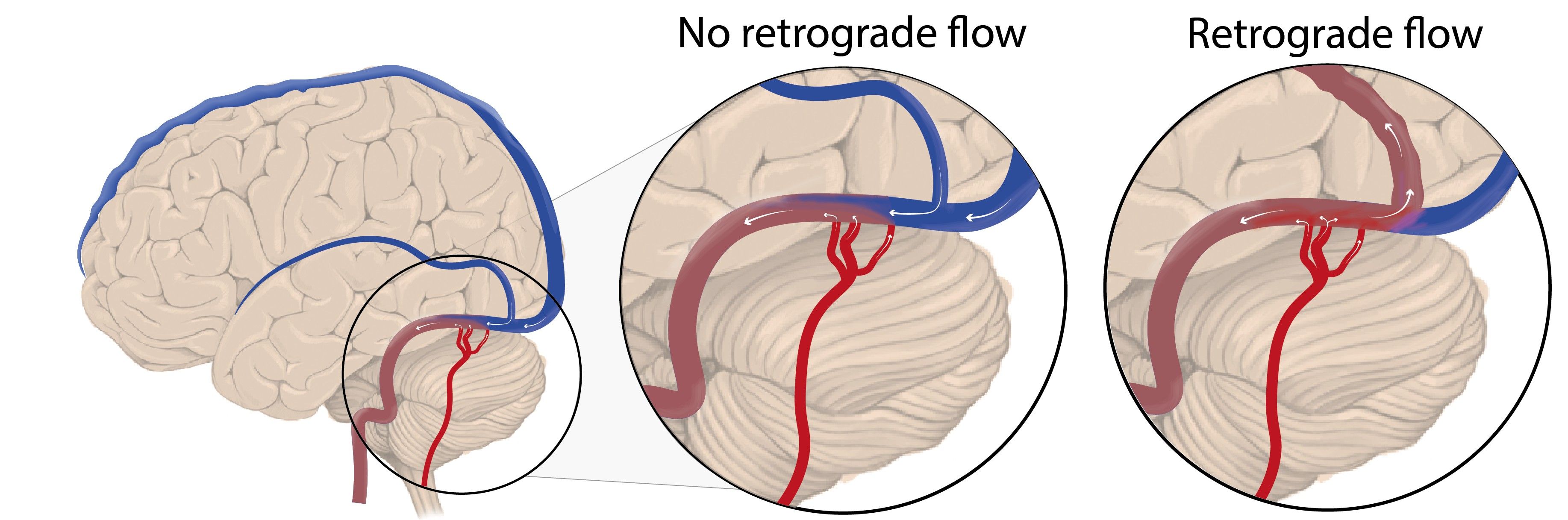
Figure 3: Dural arteriovenous fistula. On the left is a schematic diagram of the brain, with the veins shown in blue. On the right are enlarged images of dAVFs: on the left, a relatively harmless dAVF without blood pooling in the cerebral veins; on the right, a dAVF with blood pooling in the cerebral veins, which is associated with an increased risk of hemorrhage.
What symptoms can a dAVF cause?
The symptoms that occur depend primarily on the location of the dAVF and how the blood flowing through the fistula is drained. Some dAVFs cause only mild symptoms or go completely unnoticed.
Common symptoms include:
- Neurological deficits, for example
- Weakness or signs of paralysis
- Sensory disturbances
- Vision problems or speech disorders
- Balance or coordination problems
- Pulsating ringing in the ear (“tinnitus”)
- Headaches
- Vision problems with or without swelling, pain, or redness in one eye
- Epileptic Seizures
In certain cases, a life-threatening brain hemorrhage may occur. This condition may present with sudden, severe headaches, neurological deficits, or impaired consciousness, and constitutes a medical emergency.
Not every dAVF causes symptoms, and not every dAVF requires treatment. That is why a thorough evaluation is crucial.
How is a dAVF diagnosed?
Imaging tests are essential for diagnosis and assessing individual risk. They show where the fistula is located and how the blood flows out.
The following tests are used for diagnosis:
- Magnetic resonance imaging (MRI) : MRI can be used to visualize blood vessels in high resolution. In addition, MRI is particularly well-suited for visualizing changes in the brain or spinal cord caused by altered blood flow.
- Computed Tomography (CT):A CT scan is primarily used in emergency situations to quickly and reliably detect bleeding and to initiate appropriate immediate measures.
- Catheter angiography: In this procedure, a very thin tube is guided through a blood vessel in the arm or leg to the blood vessels in the brain or spinal cord. Contrast dye allows for precise visualization of the vascular connections and blood flow. This examination is particularly important for accurately assessing the risk of dAVF and planning treatment.
The imaging techniques used are tailored to each individual’s specific situation. The results are then explained to the patients in detail.
How is a dAVF treated?
Not every dAVF requires immediate treatment. Whether treatment is appropriate—and, if so, which type—depends on the individual’s risk and symptoms.
More on the Treatment of a dAVF
Cavernoma
A cavernoma is a specific malformation of the blood vessels in the brain or spinal cord. It consists of small, closely spaced, dilated vascular spaces filled with blood. Unlike in an arteriovenous malformation and a dural arteriovenous fistula, blood flows very slowly and without high pressure in these vascular spaces.
A cavernoma is relatively common andoccurs in about 1 in 200 people.⁶ Many people with this condition are unaware of their vascular malformation, since cavernomas often cause no symptoms and are discovered by chance during imaging tests.
Although blood flow is slow, the thin vessel walls of a cavernoma may be unstable. This can lead to minor or more severe bleeding, which, depending on its location in the brain, can trigger neurological symptoms. The average risk of hemorrhage from a cavernoma is approximately 2% per year.7 However, after a hemorrhage has already occurred, the risk of further hemorrhages is significantly increased, particularly for cavernomas located in the brainstem region.
Many cavernomas are congenital, but symptoms often do not appear until early or middle adulthood. These may include, among other things, epileptic seizures, neurological deficits, or headaches. Whether a cavernoma needs to be treated depends primarily on its location, the occurrence of bleeding, and the patient’s symptoms. An individualized evaluation at a specialized center is therefore crucial.
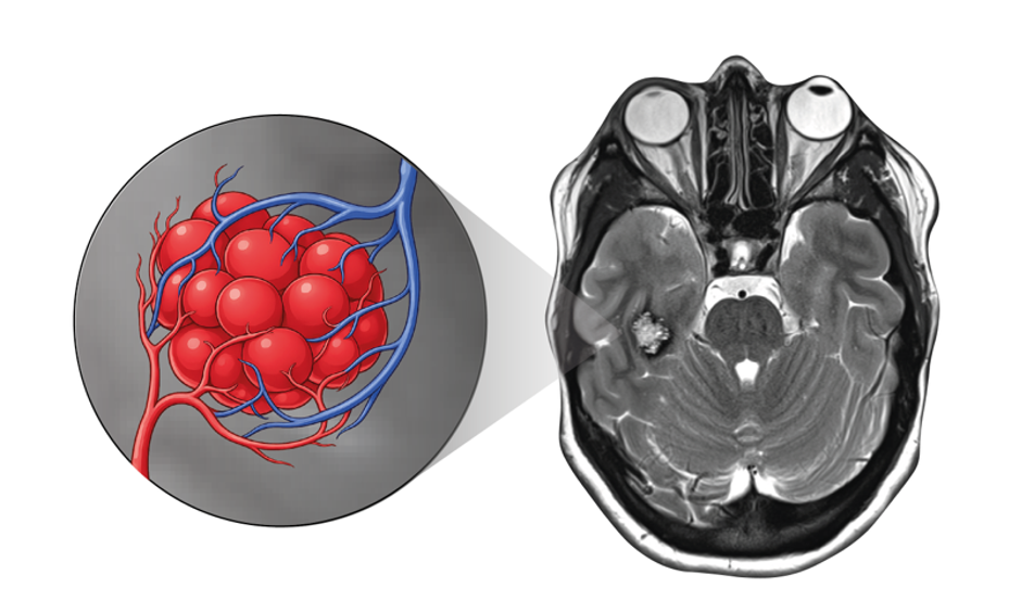
Figure 4: Cavernoma. Shown on the right is an MRI cross-sectional image depicting a typical cavernoma. The enlarged image on the left shows a schematic representation of a cavernoma, with the feeding artery shown in red and the draining vein shown in blue.
What symptoms can a cavernoma cause?
Many cavernomas remain asymptomatic for a long time and do not cause any symptoms. If symptoms occur, they usually depend on the location of the cavernoma and whether it is bleeding.
Possible symptoms include:
- Epileptic Seizures
- Neurological deficits, for example
- Weakness or paralysis
- Sensory disturbances
- Vision problems or speech disorders
- Balance or coordination problems
- Headaches, dizziness, and other symptoms
If bleeding occurs, symptoms may appear suddenly or worsen rapidly. The severity depends on the extent of the bleeding and which area of the brain is affected.
Not every cavernoma leads to bleeding or symptoms. A thorough evaluation is therefore important in order to accurately assess an individual’s risk.
How is a cavernoma diagnosed?
A cavernoma can be clearly visualized using imaging studies. The goal of the diagnostic evaluation is to assess the exact location, size, and possible signs of bleeding.
The following tests are used for diagnosis:
- Magnetic Resonance Imaging (MRI): MRI is the most important test for diagnosing a cavernoma. It can depict the typical structure of blood vessels with great precision and also reveal even minor hemorrhages.
- Computed Tomography (CT): A CT scan is performed primarily when there is suspicion of a recent hemorrhage.
Catheter angiography is generally not effective for cavernomas, as these vascular abnormalities are usually not visible during the procedure.
How is a cavernoma treated?
Not every cavernoma requires treatment. Whether treatment is advisable depends on whether symptoms are present, whether bleeding has occurred, and where the cavernoma is located.
More on the Treatment of a Cavernoma
Prevention, Early Detection, Prognosis
Targeted prevention is not possible for most vascular malformations. Since many changes do not cause any symptoms, they are often discovered by chance during other tests. A thorough evaluation by medical experts is essential for identifying high-risk forms of the disease and initiating timely treatment.
The prognosis depends on the type of vascular malformation, the individual’s risk of bleeding, the location of the malformation, and any associated symptoms. Thanks to modern diagnostic methods and targeted treatment options, risks can often be significantly reduced today, and quality of life can be maintained over the long term.
Why Choose the University Hospital of Zurich
The treatment of vascular malformations in the brain or spinal cord requires the utmost precision and extensive experience. For years, the specialists at the USZ have been making important contributions to the understanding and treatment of vascular malformations of the brain and spinal cord. At the University Hospital of Zurich, patients benefit from a combination of high levels of clinical specialization, state-of-the-art technology, and interdisciplinary collaboration—a combination that is possible in this form at only a few centers.
Interdisciplinary Case Discussions
Every vascular malformation of the brain and spinal cord is presented at a specialized conference attended by experts in neurosurgery, neuroradiology, radiation oncology, and neurology. This close collaboration enables treatment decisions that are tailored to the individual and safe.
State-of-the-art diagnostics and technology
The University Hospital of Zurich uses state-of-the-art diagnostic and treatment techniques. These technologies make it possible to visualize even the smallest vascular changes in the brain or spinal cord with great precision and to plan and carry out treatments with a high degree of safety.
All forms of therapy under one roof
The USZ offers all established treatment modalities, including microsurgery, endovascular procedures, radiation therapy, and neuro-intensive care. This allows us to combine therapeutic approaches on an individual basis and tailor them optimally to each patient’s specific condition and life situation.
- Beyersdorf, B., Schwieger, Y., Padevit, L., Kulcsar, Z., Germans, M. R., Regli, L., & Akeret, K. Anatomical phenotyping and staging of brain arteriovenous malformations: registered report. Brain Communications.
- Mast, H. et al. Risk of spontaneous hemorrhage following diagnosis of a cerebral arteriovenous malformation. Lancet 350, 1065–1068 (1997).
- Gross, B. A. & Du, R. Rate of rebleeding from arteriovenous malformations in the first year after rupture. J. Clin. Neurosci. 19, 1087–1088 (2012).
- Baltsavias, G., Roth, P., & Valavanis, A. Cranial dural arteriovenous shunts. Part 3. Classification Based on Leptomeningeal Venous Drainage. Neurosurg. Rev. 38, 273–81; discussion 281 (2015).
- van Dijk, J. M. C., terBrugge, K. G., Willinsky, R. A., & Wallace, M. C. Clinical course of cranial dural arteriovenous fistulas with long-term persistent cortical venous reflux. Stroke 33, 1233–1236 (2002).
- Flemming, K. D. et al. Population-based prevalence of cerebral cavernous malformations in older adults: Mayo Clinic Study of Aging. JAMA Neurol. 74, 801–805 (2017).
- Kashefiolasl, S. et al. A benchmark approach to hemorrhage risk management in cavernous malformations. Neurology 90, e856–e863 (2018).