Using cardiac MRI, the effects of HCM and storage diseases on the structure (hypertrophy or fibrosis) and function of the heart muscle can be examined in detail.
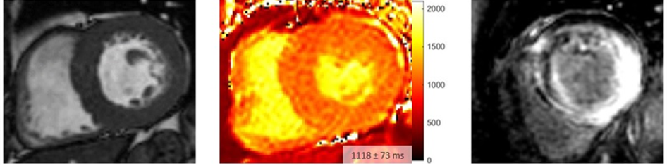
Typical image of cardiac amyloidosis in cardiac MRI with concentric hypertrophy (left), significantly increased T1 mapping (center) and pronounced myocardial late gadolinium enhancement (LGE, right).
In addition, the magnetic relaxation properties of the heart muscle change in a characteristic way in storage diseases. A particular strength of cardiac MRI is its ability to quantify these relaxation characteristics (T1, T2 and T2*) and thus provide important information for diagnosis, particularly in the early stages of the disease.
When diagnosing patients with storage diseases, the radiology department works closely with an interdisciplinary team of specialists to ensure optimal care(amyloidosis network).
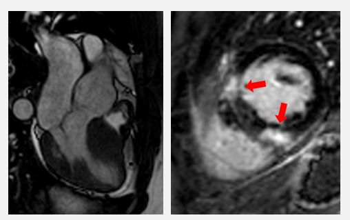
Cardiac MRI of a patient with HCM. There is clear hypertrophy of the septum (left) and typical fibrosis, emphasized at the base of the right ventricle (right, red arrows).