Cardiovascular Imaging Group
The Cardiovascular Imaging Research Group is an interdisciplinary research group in the Institute of Diagnostic and Interventional Radiology at the University Hospital Zurich, Switzerland. The group includes faculty members, senior and junior radiologists, postdoctoral fellows, research staff, and medical students and has the aim to pursue teaching and research in multi-modality cardiovascular imaging.
In close collaboration with various clinical partners at the University Hospital Zurich such as the Department of Cardiology and the Division of Cardiovascular Surgery as well as the Institute for Biomedical Engineering, an institution of the Swiss Federal Institute of Technology (ETH Zurich) the Cardiovascular Imaging Research Group aims to integrate research into clinical routine and to build collaborations between scientists, physicians, and radiologists establishing a strong resource for imaging-based research at the University of Zurich.
Main interests of the cardiovascular imaging research group include the following modalities and research topics:
Computed Tomography (CT)
- Coronary Heart Disease
- Implementation and assessment of image quality of new CT protocols
- Ultra low dose CT coronary angiography protocols
- Dosimetry
- Myocardial Perfusion Imaging
- Imaging of Cardiac Valve Anatomy and Function
Magnetic Resonance Imaging (MRI)
- Imaging of Myocardial Ischemia and Infarcts
- Imaging of Cardiomyopathy
- Imaging in Grown-up Congenital Heart Disease
- Cardiac Anatomy and Tissue Characterization
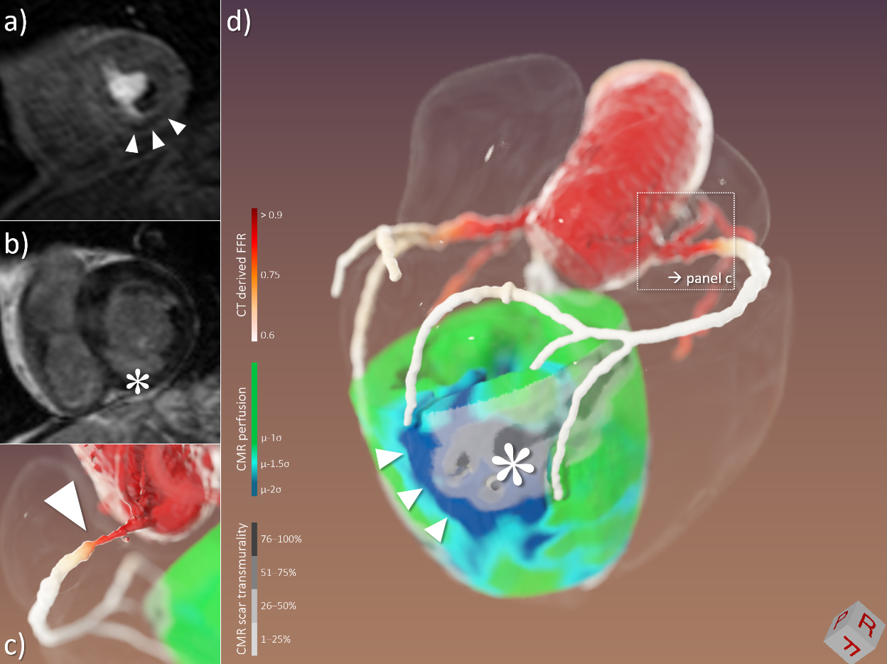
3D image fusion combining information from CT coronary angiography, CT derived FFR, CMR stress perfusion imaging, and CMR late gadolinium enhancement. Data from a 59-year old male patient with severe three-vessel disease are shown. Conventional 2D images of CT and CMR datasets (a: CMR stress perfusion, b: CMR late gadolinium enhancement) were post-processed, co-registered, color-coded, and rendered in a three-dimensional fashion. In CT coronary angiography, a subtotal proximal stenosis of the right coronary artery was found (c: 3D rendering, also note the associated drop of CT derived FFR value), which resulted in an inferior/inferolateral perfusion deficit (arrowheads in a and d) as well as severe, partly transmural scar (asterisk in b and d).


Technical Resources
- MRI (different vendors ; 1 x 1.5T; 3 x 3.0T; 1 x 7.0T)
- CT: dual-source 128-slice CT (Somatom Definition Flash), dual-source 192-slice CT (Somatom Force), single-source 64-slice CT (Somatom Definition AS)
- PET-CT and PET-MR
- 3D Lab